 Movie
Movie Controller

[English] 日本語
 Yorodumi
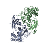
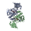
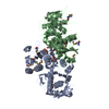
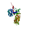
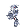
- PDB-5kye: Crystal structure of USP7 catalytic domain [H294E] mutant in comp... -
+ Open data

- Basic information
| Entry | Database: PDB / ID: 5kye | ||||||
|---|---|---|---|---|---|---|---|
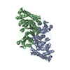
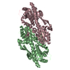
| Title | Crystal structure of USP7 catalytic domain [H294E] mutant in complex with ubiquitin | ||||||
 Components Components |
| ||||||
 Keywords Keywords |  HYDROLASE / USP7 catalytic domain / deubiquitinase / H294E mutant / ubiquitin HYDROLASE / USP7 catalytic domain / deubiquitinase / H294E mutant / ubiquitin | ||||||
| Function / homology |  Function and homology information Function and homology informationregulation of telomere capping / : / monoubiquitinated protein deubiquitination / regulation of retrograde transport, endosome to Golgi / deubiquitinase activity / negative regulation of gene expression via chromosomal CpG island methylation / regulation of DNA-binding transcription factor activity / hypothalamus gonadotrophin-releasing hormone neuron development / K48-linked deubiquitinase activity / female meiosis I ...regulation of telomere capping / : / monoubiquitinated protein deubiquitination / regulation of retrograde transport, endosome to Golgi / deubiquitinase activity / negative regulation of gene expression via chromosomal CpG island methylation / regulation of DNA-binding transcription factor activity / hypothalamus gonadotrophin-releasing hormone neuron development / K48-linked deubiquitinase activity / female meiosis I / positive regulation of protein monoubiquitination / mitochondrion transport along microtubule / symbiont-mediated disruption of host cell PML body / fat pad development / female gonad development / negative regulation of NF-kappaB transcription factor activity / seminiferous tubule development / protein deubiquitination / male meiosis I / positive regulation of intrinsic apoptotic signaling pathway by p53 class mediator / negative regulation of proteasomal ubiquitin-dependent protein catabolic process / transcription-coupled nucleotide-excision repair / negative regulation of gluconeogenesis / energy homeostasis / regulation of neuron apoptotic process / regulation of proteasomal protein catabolic process / Maturation of protein E / Maturation of protein E / ER Quality Control Compartment (ERQC) / Myoclonic epilepsy of Lafora / negative regulation of TORC1 signaling / FLT3 signaling by CBL mutants / Prevention of phagosomal-lysosomal fusion / IRAK2 mediated activation of TAK1 complex / Alpha-protein kinase 1 signaling pathway / Glycogen synthesis / IRAK1 recruits IKK complex / IRAK1 recruits IKK complex upon TLR7/8 or 9 stimulation / Membrane binding and targetting of GAG proteins / Constitutive Signaling by NOTCH1 HD Domain Mutants / Endosomal Sorting Complex Required For Transport (ESCRT) / NOTCH2 Activation and Transmission of Signal to the Nucleus / IRAK2 mediated activation of TAK1 complex upon TLR7/8 or 9 stimulation / PTK6 Regulates RTKs and Their Effectors AKT1 and DOK1 / Negative regulation of FLT3 / Regulation of FZD by ubiquitination / TICAM1,TRAF6-dependent induction of TAK1 complex / TICAM1-dependent activation of IRF3/IRF7 / APC/C:Cdc20 mediated degradation of Cyclin B / p75NTR recruits signalling complexes / Downregulation of ERBB4 signaling / TRAF6 mediated IRF7 activation in TLR7/8 or 9 signaling / APC-Cdc20 mediated degradation of Nek2A / PINK1-PRKN Mediated Mitophagy / TRAF6-mediated induction of TAK1 complex within TLR4 complex / Pexophagy / InlA-mediated entry of Listeria monocytogenes into host cells / Regulation of innate immune responses to cytosolic DNA / VLDLR internalisation and degradation / Downregulation of ERBB2:ERBB3 signaling / NRIF signals cell death from the nucleus / Activated NOTCH1 Transmits Signal to the Nucleus / NF-kB is activated and signals survival / Regulation of PTEN localization / Regulation of BACH1 activity / Translesion synthesis by REV1 / Synthesis of active ubiquitin: roles of E1 and E2 enzymes / Translesion synthesis by POLK / MAP3K8 (TPL2)-dependent MAPK1/3 activation / TICAM1, RIP1-mediated IKK complex recruitment / Gap-filling DNA repair synthesis and ligation in GG-NER / Downregulation of TGF-beta receptor signaling / Translesion synthesis by POLI / Josephin domain DUBs / Activation of IRF3, IRF7 mediated by TBK1, IKKε (IKBKE) / Regulation of activated PAK-2p34 by proteasome mediated degradation / neuron projection morphogenesis / InlB-mediated entry of Listeria monocytogenes into host cell / IKK complex recruitment mediated by RIP1 / JNK (c-Jun kinases) phosphorylation and activation mediated by activated human TAK1 / regulation of mitochondrial membrane potential / TGF-beta receptor signaling in EMT (epithelial to mesenchymal transition) / N-glycan trimming in the ER and Calnexin/Calreticulin cycle / Autodegradation of Cdh1 by Cdh1:APC/C / TNFR1-induced NF-kappa-B signaling pathway / APC/C:Cdc20 mediated degradation of Securin / regulation of signal transduction by p53 class mediator / positive regulation of protein ubiquitination / Asymmetric localization of PCP proteins / TCF dependent signaling in response to WNT / SCF-beta-TrCP mediated degradation of Emi1 / NIK-->noncanonical NF-kB signaling / Regulation of NF-kappa B signaling / Ubiquitin-dependent degradation of Cyclin D / AUF1 (hnRNP D0) binds and destabilizes mRNA / NOTCH3 Activation and Transmission of Signal to the Nucleus / TNFR2 non-canonical NF-kB pathway / activated TAK1 mediates p38 MAPK activation / Assembly of the pre-replicative complex / Vpu mediated degradation of CD4Similarity search - Function | ||||||
| Biological species |  Homo sapiens (human) Homo sapiens (human) | ||||||
| Method | X-RAY DIFFRACTION / SYNCHROTRON / MOLECULAR REPLACEMENT / molecular replacement / Resolution: 1.97 Å | ||||||
 Authors Authors | Rouge, L. / Ozen, A. | ||||||
 Citation Citation | Journal: Structure / Year: 2018 Title: Selectively Modulating Conformational States of USP7 Catalytic Domain for Activation. Authors: Ozen, A. / Rouge, L. / Bashore, C. / Hearn, B.R. / Skelton, N.J. / Dueber, E.C. | ||||||
| History |
|
- Structure visualization
| Structure viewer | Molecule: MolmilJmol/JSmol |
|---|
- Downloads & links
-Download
| PDBx/mmCIF format | 5kye.cif.gz | 186.1 KB | Display | PDBx/mmCIF format |
|---|---|---|---|---|
| PDB format | pdb5kye.ent.gz | 146.1 KB | Display | PDB format |
| PDBx/mmJSON format | 5kye.json.gz | Tree view | PDBx/mmJSON format | |
| Others |  Other downloads Other downloads |
-Validation report
| Arichive directory | https://data.pdbj.org/pub/pdb/validation_reports/ky/5kyeftp://data.pdbj.org/pub/pdb/validation_reports/ky/5kye | HTTPS FTP |
|---|
-Related structure data
| Related structure data |  5kybC 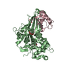 5kycC 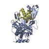 5kydC 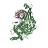 5kyfC 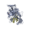 1nbfS C: citing same article ( S: Starting model for refinement |
|---|---|
| Similar structure data |

-Links
 PDBj
PDBj


- Assembly
| Deposited unit | 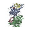
| ||||||||
|---|---|---|---|---|---|---|---|---|---|
| 1 | 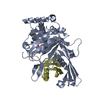
| ||||||||
| 2 | 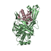
| ||||||||
| Unit cell |
|
-Components
| #1: Protein | Mass: 40578.848 Da / Num. of mol.: 2 / Fragment: UNP residues 192-538 / Mutation: H294E Source method: isolated from a genetically manipulated source Source: (gene. exp.) Homo sapiens (human) / Gene: USP7, HAUSP / Production host:  Escherichia coli (E. coli) / References: UniProt: Q93009, ubiquitinyl hydrolase 1 Escherichia coli (E. coli) / References: UniProt: Q93009, ubiquitinyl hydrolase 1#2: Protein | Mass: 8576.831 Da / Num. of mol.: 2 / Fragment: UNP residues 1-76 Source method: isolated from a genetically manipulated source Source: (gene. exp.) Homo sapiens (human) / Gene: UBB / Production host: Escherichia coli (E. coli) / References: UniProt: P0CG47#3: Chemical | ChemComp-EPE / | HEPES  Mass: 238.305 Da / Num. of mol.: 1 / Source method: obtained synthetically / Formula: C8H18N2O4S / Comment: pH buffer*YM Mass: 238.305 Da / Num. of mol.: 1 / Source method: obtained synthetically / Formula: C8H18N2O4S / Comment: pH buffer*YM#4: Water | ChemComp-HOH / | Water Mass: 18.015 Da / Num. of mol.: 390 / Source method: isolated from a natural source / Formula: H2O Mass: 18.015 Da / Num. of mol.: 390 / Source method: isolated from a natural source / Formula: H2O |
|---|
-Experimental details
-Experiment
| Experiment | Method: X-RAY DIFFRACTION / Number of used crystals: 1 |
|---|
- Sample preparation
| Crystal | Density Matthews: 2.11 Å3/Da / Density % sol: 41.74 % |
|---|---|
| Crystal grow | Temperature: 292 K / Method: vapor diffusion, hanging drop / pH: 7.5 Details: 0.1M HEPES pH 7.5, 25% PEG3350, 0.2M Ammonium acetate |
-Data collection
| Diffraction | Mean temperature: 100 K | ||||||||||||||||||
|---|---|---|---|---|---|---|---|---|---|---|---|---|---|---|---|---|---|---|---|
| Diffraction source | Source: SYNCHROTRON / Site: ALS  / Beamline: 5.0.2 / Wavelength: 0.98 Å / Beamline: 5.0.2 / Wavelength: 0.98 Å | ||||||||||||||||||
| Detector | Type: ADSC QUANTUM 315r / Detector: CCD / Date: Feb 24, 2016 | ||||||||||||||||||
| Radiation | Protocol: SINGLE WAVELENGTH / Monochromatic (M) / Laue (L): M / Scattering type: x-ray | ||||||||||||||||||
| Radiation wavelength | Wavelength: 0.98 Å / Relative weight: 1 | ||||||||||||||||||
| Reflection | Resolution: 1.97→53.892 Å / Num. obs: 57873 / % possible obs: 100 % / Redundancy: 6 % / Biso Wilson estimate: 29.36 Å2 / CC1/2: 0.999 / Rmerge(I) obs: 0.08 / Net I/σ(I): 12.4 | ||||||||||||||||||
| Reflection shell |
|
-Phasing
| Phasing | Method: molecular replacement |
|---|
- Processing
| Software |
| ||||||||||||||||||||||||
|---|---|---|---|---|---|---|---|---|---|---|---|---|---|---|---|---|---|---|---|---|---|---|---|---|---|
| Refinement | Method to determine structure: MOLECULAR REPLACEMENT Starting model: 1NBF Resolution: 1.97→53.892 Å / SU ML: 0.22 / Cross valid method: NONE / σ(F): 1.34 / Phase error: 23.54 / Stereochemistry target values: ML
| ||||||||||||||||||||||||
| Solvent computation | Shrinkage radii: 0.9 Å / VDW probe radii: 1.11 Å / Solvent model: FLAT BULK SOLVENT MODEL | ||||||||||||||||||||||||
| Displacement parameters | Biso max: 206.73 Å2 / Biso mean: 39.2332 Å2 / Biso min: 17.19 Å2 | ||||||||||||||||||||||||
| Refinement step | Cycle: final / Resolution: 1.97→53.892 Å
|
