 Movie
Movie Controller


+ Open data

- Basic information
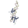
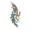
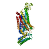
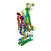
| Entry | Database: PDB / ID: 1ay9 | ||||||
|---|---|---|---|---|---|---|---|
| Title | WILD-TYPE UMUD' FROM E. COLI | ||||||
 Components Components | UMUD PROTEIN | ||||||
 Keywords Keywords |  MUTAGENESIS PROTEIN / DNA REPAIR / HYDROLASE MUTAGENESIS PROTEIN / DNA REPAIR / HYDROLASE | ||||||
| Function / homology |  Function and homology informationDNA polymerase V complex / SOS response / Hydrolases; Acting on peptide bonds (peptidases); Serine endopeptidases / ATP-dependent activity, acting on DNA / translesion synthesis / serine-type peptidase activity / single-stranded DNA binding / nucleic acid binding / DNA-directed DNA polymerase activity / DNA repair ...DNA polymerase V complex / SOS response / Hydrolases; Acting on peptide bonds (peptidases); Serine endopeptidases / ATP-dependent activity, acting on DNA / translesion synthesis / serine-type peptidase activity / single-stranded DNA binding / nucleic acid binding / DNA-directed DNA polymerase activity / DNA repair / DNA damage response / regulation of DNA-templated transcription / proteolysis / DNA binding / identical protein binding Function and homology informationDNA polymerase V complex / SOS response / Hydrolases; Acting on peptide bonds (peptidases); Serine endopeptidases / ATP-dependent activity, acting on DNA / translesion synthesis / serine-type peptidase activity / single-stranded DNA binding / nucleic acid binding / DNA-directed DNA polymerase activity / DNA repair ...DNA polymerase V complex / SOS response / Hydrolases; Acting on peptide bonds (peptidases); Serine endopeptidases / ATP-dependent activity, acting on DNA / translesion synthesis / serine-type peptidase activity / single-stranded DNA binding / nucleic acid binding / DNA-directed DNA polymerase activity / DNA repair / DNA damage response / regulation of DNA-templated transcription / proteolysis / DNA binding / identical protein bindingSimilarity search - Function | ||||||
| Biological species |  Escherichia coli (E. coli) Escherichia coli (E. coli) | ||||||
| Method | X-RAY DIFFRACTION / SYNCHROTRON / MOLECULAR REPLACEMENT / Resolution: 3 Å | ||||||
 Authors Authors | Peat, T.S. / Frank, E.G. / Mcdonald, J.P. / Levine, A.S. / Woodgate, R. / Hendrickson, W.A. | ||||||
 Citation Citation | Journal: Structure / Year: 1996 Title: The UmuD' protein filament and its potential role in damage induced mutagenesis. Authors: Peat, T.S. / Frank, E.G. / McDonald, J.P. / Levine, A.S. / Woodgate, R. / Hendrickson, W.A. #1: Journal: Nature / Year: 1996Title: Structure of the UmuD' Protein and its Regulation in Response to DNA Damage Authors: Peat, T.S. / Frank, E.G. / Mcdonald, J.P. / Levine, A.S. / Woodgate, R. / Hendrickson, W.A. | ||||||
| History |
|
- Structure visualization
| Structure viewer | Molecule: MolmilJmol/JSmol |
|---|
- Downloads & links
-Download
| PDBx/mmCIF format | 1ay9.cif.gz | 49.6 KB | Display | PDBx/mmCIF format |
|---|---|---|---|---|
| PDB format | pdb1ay9.ent.gz | 35.9 KB | Display | PDB format |
| PDBx/mmJSON format | 1ay9.json.gz | Tree view | PDBx/mmJSON format | |
| Others |  Other downloads Other downloads |
-Validation report
| Arichive directory | https://data.pdbj.org/pub/pdb/validation_reports/ay/1ay9ftp://data.pdbj.org/pub/pdb/validation_reports/ay/1ay9 | HTTPS FTP |
|---|
-Related structure data
| Related structure data |  1umuS S: Starting model for refinement |
|---|---|
| Similar structure data |


-Links
 PDBj
PDBj- Assembly
| Deposited unit | 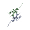
| ||||||||
|---|---|---|---|---|---|---|---|---|---|
| 1 | 
| ||||||||
| 2 | 
| ||||||||
| Unit cell |
| ||||||||
| Noncrystallographic symmetry (NCS) | NCS oper: (Code: given Matrix: (0.341469, 0.939004, -0.040869), Vector : |
-Components
| #1: Protein | Mass: 11667.270 Da / Num. of mol.: 2 / Source method: isolated from a natural source / Source: (natural) Escherichia coli (E. coli)References: UniProt: P04153, UniProt: P0AG11*PLUS, Hydrolases; Acting on peptide bonds (peptidases); Serine endopeptidases#2: Water | ChemComp-HOH / | Water Mass: 18.015 Da / Num. of mol.: 36 / Source method: isolated from a natural source / Formula: H2O Mass: 18.015 Da / Num. of mol.: 36 / Source method: isolated from a natural source / Formula: H2O |
|---|
-Experimental details
-Experiment
| Experiment | Method: X-RAY DIFFRACTION / Number of used crystals: 1 |
|---|
- Sample preparation
| Crystal | Density Matthews: 2.48 Å3/Da / Density % sol: 47 % | ||||||||||||||||||||||||||||||||||||||||||
|---|---|---|---|---|---|---|---|---|---|---|---|---|---|---|---|---|---|---|---|---|---|---|---|---|---|---|---|---|---|---|---|---|---|---|---|---|---|---|---|---|---|---|---|
| Crystal grow | pH: 6 / Details: 100MM CACODYLATE BUFFER PH 6.0 | ||||||||||||||||||||||||||||||||||||||||||
| Crystal grow | *PLUS Temperature: 20 ℃ / pH: 5.8 / Method: vapor diffusion, hanging drop | ||||||||||||||||||||||||||||||||||||||||||
| Components of the solutions | *PLUS
|
-Data collection
| Diffraction | Mean temperature: 277 K |
|---|---|
| Diffraction source | Source: SYNCHROTRON / Site: CHESS  / Beamline: F1 / Wavelength: 0.918 / Beamline: F1 / Wavelength: 0.918 |
| Detector | Type: FUJI / Detector: IMAGE PLATE / Date: Dec 1, 1990 |
| Radiation | Monochromator: SINGLE CRYSTAL / Monochromatic (M) / Laue (L): M / Scattering type: x-ray |
| Radiation wavelength | Wavelength: 0.918 Å / Relative weight: 1 |
| Reflection | Resolution: 3→15 Å / Num. obs: 4912 / % possible obs: 90 % / Observed criterion σ(I): 2 / Redundancy: 3.2 % |
- Processing
| Software |
| ||||||||||||||||||||||||||||||||||||||||||||||||||||||||||||
|---|---|---|---|---|---|---|---|---|---|---|---|---|---|---|---|---|---|---|---|---|---|---|---|---|---|---|---|---|---|---|---|---|---|---|---|---|---|---|---|---|---|---|---|---|---|---|---|---|---|---|---|---|---|---|---|---|---|---|---|---|---|
| Refinement | Method to determine structure: MOLECULAR REPLACEMENT Starting model: PDB ENTRY 1UMU Resolution: 3→15 Å / Cross valid method: THROUGHOUT / σ(F): 2
| ||||||||||||||||||||||||||||||||||||||||||||||||||||||||||||
| Displacement parameters | Biso mean: 10.7 Å2 | ||||||||||||||||||||||||||||||||||||||||||||||||||||||||||||
| Refinement step | Cycle: LAST / Resolution: 3→15 Å
| ||||||||||||||||||||||||||||||||||||||||||||||||||||||||||||
| Refine LS restraints |
| ||||||||||||||||||||||||||||||||||||||||||||||||||||||||||||
| Refine LS restraints NCS | NCS model details: RESTRAINTS | ||||||||||||||||||||||||||||||||||||||||||||||||||||||||||||
| LS refinement shell | Resolution: 3→3.11 Å / Total num. of bins used: 10
| ||||||||||||||||||||||||||||||||||||||||||||||||||||||||||||
| Xplor file |
|
