 Movie
Movie Controller

[English] 日本語
 Yorodumi
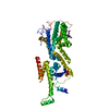
- PDB-6ytz: Crystal structure of Malus domestica Double Bond Reductase (MdDBR... -
+ Open data

- Basic information
| Entry | Database: PDB / ID: 6ytz | ||||||
|---|---|---|---|---|---|---|---|
| Title | Crystal structure of Malus domestica Double Bond Reductase (MdDBR) in complex with NADPH | ||||||
 Components Components | Double Bond Reductase | ||||||
 Keywords Keywords | BIOSYNTHETIC PROTEIN / phenylpropanoid pathway / double bond reductase / Malus domestica | ||||||
| Function / homology | NADP NICOTINAMIDE-ADENINE-DINUCLEOTIDE PHOSPHATE / R-1,2-PROPANEDIOL Function and homology information Function and homology information | ||||||
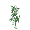
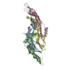
| Biological species |  | ||||||
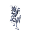
| Method |  X-RAY DIFFRACTION / SYNCHROTRON / MOLECULAR REPLACEMENT / molecular replacement / Resolution: 1.4 Å X-RAY DIFFRACTION / SYNCHROTRON / MOLECULAR REPLACEMENT / molecular replacement / Resolution: 1.4 Å | ||||||
 Authors Authors | Caliandro, R. / Polsinelli, I. / Demitri, N. / Benini, S. | ||||||
 Citation Citation | Journal: Int.J.Biol.Macromol. / Year: 2021 Title: The structural and functional characterization of Malus domestica double bond reductase MdDBR provides insights towards the identification of its substrates. Authors: Caliandro, R. / Polsinelli, I. / Demitri, N. / Musiani, F. / Martens, S. / Benini, S. | ||||||
| History |
|
- Structure visualization
| Structure viewer | Molecule: MolmilJmol/JSmol |
|---|
- Downloads & links
-Download
| PDBx/mmCIF format | 6ytz.cif.gz | 166.7 KB | Display | PDBx/mmCIF format |
|---|---|---|---|---|
| PDB format | pdb6ytz.ent.gz | 129.9 KB | Display | PDB format |
| PDBx/mmJSON format | 6ytz.json.gz | Tree view | PDBx/mmJSON format | |
| Others |  Other downloads Other downloads |
-Validation report
| Summary document | 6ytz_validation.pdf.gz | 748.4 KB | Display | wwPDB validaton report |
|---|---|---|---|---|
| Full document | 6ytz_full_validation.pdf.gz | 751.8 KB | Display | |
| Data in XML | 6ytz_validation.xml.gz | 18.4 KB | Display | |
| Data in CIF | 6ytz_validation.cif.gz | 27.5 KB | Display | |
| Arichive directory | https://data.pdbj.org/pub/pdb/validation_reports/yt/6ytzftp://data.pdbj.org/pub/pdb/validation_reports/yt/6ytz | HTTPS FTP |
-Related structure data
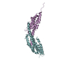
| Related structure data | 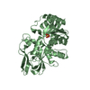 6ysbSC 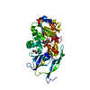 6yuxC S: Starting model for refinement C: citing same article ( |
|---|---|
| Similar structure data |


-Links
 PDBj
PDBj- Assembly
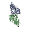
| Deposited unit | 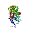
| |||||||||||||||
|---|---|---|---|---|---|---|---|---|---|---|---|---|---|---|---|---|
| 1 | 
| |||||||||||||||
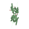
| Unit cell |
| |||||||||||||||
| Components on special symmetry positions |
|
-Components
-Protein , 1 types, 1 molecules A
| #1: Protein | Mass: 38484.410 Da / Num. of mol.: 1 Source method: isolated from a genetically manipulated source Source: (gene. exp.)  |
|---|
-Non-polymers , 6 types, 292 molecules 


| #2: Chemical | ChemComp-NAP /  Mass: 743.405 Da / Num. of mol.: 1 / Source method: obtained synthetically / Formula: C21H28N7O17P3 Mass: 743.405 Da / Num. of mol.: 1 / Source method: obtained synthetically / Formula: C21H28N7O17P3 |
|---|---|
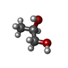
| #3: Chemical | ChemComp-MPD / ( Mass: 118.174 Da / Num. of mol.: 1 / Source method: obtained synthetically / Formula: C6H14O2 / Comment: precipitant*YM Mass: 118.174 Da / Num. of mol.: 1 / Source method: obtained synthetically / Formula: C6H14O2 / Comment: precipitant*YM |
| #4: Chemical | ChemComp-EDO /  Mass: 62.068 Da / Num. of mol.: 1 / Source method: obtained synthetically / Formula: C2H6O2 Mass: 62.068 Da / Num. of mol.: 1 / Source method: obtained synthetically / Formula: C2H6O2 |
| #5: Chemical | ChemComp-PGR /  Mass: 76.094 Da / Num. of mol.: 1 / Source method: isolated from a natural source / Formula: C3H8O2 Mass: 76.094 Da / Num. of mol.: 1 / Source method: isolated from a natural source / Formula: C3H8O2 |
| #6: Chemical | ChemComp-NA /  Mass: 22.990 Da / Num. of mol.: 1 / Source method: obtained synthetically / Formula: Na Mass: 22.990 Da / Num. of mol.: 1 / Source method: obtained synthetically / Formula: Na |
| #7: Water | ChemComp-HOH / Mass: 18.015 Da / Num. of mol.: 287 / Source method: isolated from a natural source / Formula: H2O |
-Details
| Has ligand of interest | N |
|---|
-Experimental details
-Experiment
| Experiment | Method: X-RAY DIFFRACTION / Number of used crystals: 1 |
|---|
- Sample preparation
| Crystal | Density Matthews: 2.21 Å3/Da / Density % sol: 44.23 % |
|---|---|
| Crystal grow | Temperature: 293.15 K / Method: vapor diffusion, hanging drop Details: 12.5% PEG 1000, 12.5% PEG 3350, 12.5% MPD, 0.03 M magnesium chloride, 0.03 M calcium chloride, 0.03 M sodium chloride, 0.03 M sodium bromide, 0.03 M sodium iodide in 0.1 M MES/imidazole pH 6.5. |
-Data collection
| Diffraction | Mean temperature: 100 K / Serial crystal experiment: N |
|---|---|
| Diffraction source | Source: SYNCHROTRON / Site: ELETTRA  / Beamline: 5.2R / Wavelength: 1 Å / Beamline: 5.2R / Wavelength: 1 Å |
| Detector | Type: DECTRIS PILATUS 2M / Detector: PIXEL / Date: Dec 4, 2018 |
| Radiation | Protocol: SINGLE WAVELENGTH / Monochromatic (M) / Laue (L): M / Scattering type: x-ray |
| Radiation wavelength | Wavelength: 1 Å / Relative weight: 1 |
| Reflection | Resolution: 1.4→48.5 Å / Num. obs: 67744 / % possible obs: 100 % / Redundancy: 12.47 % / CC1/2: 1 / Net I/σ(I): 15.9 |
| Reflection shell | Resolution: 1.4→1.42 Å / Num. unique obs: 3301 / CC1/2: 0.517 |
-Phasing
| Phasing | Method: molecular replacement |
|---|
- Processing
| Software |
| |||||||||||||||||||||||||||||||||||||||||||||||||||||||||||||||||
|---|---|---|---|---|---|---|---|---|---|---|---|---|---|---|---|---|---|---|---|---|---|---|---|---|---|---|---|---|---|---|---|---|---|---|---|---|---|---|---|---|---|---|---|---|---|---|---|---|---|---|---|---|---|---|---|---|---|---|---|---|---|---|---|---|---|---|
| Refinement | Method to determine structure: MOLECULAR REPLACEMENT Starting model: 6YSB Resolution: 1.4→48.44 Å / Cor.coef. Fo:Fc: 0.985 / Cor.coef. Fo:Fc free: 0.976 / SU B: 2.277 / SU ML: 0.038 / Cross valid method: THROUGHOUT / σ(F): 0 / ESU R: 0.05 / ESU R Free: 0.052 / Stereochemistry target values: MAXIMUM LIKELIHOOD Details: HYDROGENS HAVE BEEN ADDED IN THE RIDING POSITIONS U VALUES : REFINED INDIVIDUALLY
| |||||||||||||||||||||||||||||||||||||||||||||||||||||||||||||||||
| Solvent computation | Ion probe radii: 0.8 Å / Shrinkage radii: 0.8 Å / VDW probe radii: 1.2 Å / Solvent model: MASK | |||||||||||||||||||||||||||||||||||||||||||||||||||||||||||||||||
| Displacement parameters | Biso max: 133.2 Å2 / Biso mean: 25.337 Å2 / Biso min: 13.7 Å2
| |||||||||||||||||||||||||||||||||||||||||||||||||||||||||||||||||
| Refinement step | Cycle: final / Resolution: 1.4→48.44 Å
| |||||||||||||||||||||||||||||||||||||||||||||||||||||||||||||||||
| Refine LS restraints |
| |||||||||||||||||||||||||||||||||||||||||||||||||||||||||||||||||
| LS refinement shell | Resolution: 1.4→1.436 Å / Rfactor Rfree error: 0 / Total num. of bins used: 20
|
