 Movie
Movie Controller

[English] 日本語
 Yorodumi
- PDB-1ajs: REFINEMENT AND COMPARISON OF THE CRYSTAL STRUCTURES OF PIG CYTOSO... -
+ Open data

- Basic information
| Entry | Database: PDB / ID: 1ajs | ||||||
|---|---|---|---|---|---|---|---|
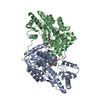
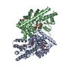
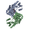
| Title | REFINEMENT AND COMPARISON OF THE CRYSTAL STRUCTURES OF PIG CYTOSOLIC ASPARTATE AMINOTRANSFERASE AND ITS COMPLEX WITH 2-METHYLASPARTATE | ||||||
 Components Components | (ASPARTATE AMINOTRANSFERASE Aspartate transaminase) x 2 Aspartate transaminase) x 2 | ||||||
 Keywords Keywords | AMINOTRANSFERASE / CYTOSOLIC ASPARTATE AMINOTRANSFERASE / PIG / IN THE PRESENCE OF LIGAND 2-METHYLASPARTATE | ||||||
| Function / homology |  Function and homology information Function and homology informationAspartate and asparagine metabolism / phosphatidylserine decarboxylase activity / glutamate catabolic process to aspartate / glutamate catabolic process to 2-oxoglutarate / cysteine transaminase / L-cysteine transaminase activity / aspartate biosynthetic process / glycerol biosynthetic process / aspartate metabolic process / Gluconeogenesis ...Aspartate and asparagine metabolism / phosphatidylserine decarboxylase activity / glutamate catabolic process to aspartate / glutamate catabolic process to 2-oxoglutarate / cysteine transaminase / L-cysteine transaminase activity / aspartate biosynthetic process / glycerol biosynthetic process / aspartate metabolic process / Gluconeogenesis / glutamate metabolic process / aspartate catabolic process / 2-oxoglutarate metabolic process / aspartate transaminase / oxaloacetate metabolic process / L-aspartate:2-oxoglutarate aminotransferase activity / fatty acid homeostasis / response to glucocorticoid / Notch signaling pathway / gluconeogenesis / cellular response to insulin stimulus / pyridoxal phosphate binding / cytosolSimilarity search - Function | ||||||
| Biological species |  Sus scrofa (pig) Sus scrofa (pig) | ||||||
| Method | X-RAY DIFFRACTION / ISOMORPHOUS DIFFERENCE MAP / Resolution: 1.6 Å | ||||||
 Authors Authors | Rhee, S. / Silva, M.M. / Hyde, C.C. / Rogers, P.H. / Metzler, C.M. / Metzler, D.E. / Arnone, A. | ||||||
 Citation Citation | Journal: J.Biol.Chem. / Year: 1997 Title: Refinement and comparisons of the crystal structures of pig cytosolic aspartate aminotransferase and its complex with 2-methylaspartate. Authors: Rhee, S. / Silva, M.M. / Hyde, C.C. / Rogers, P.H. / Metzler, C.M. / Metzler, D.E. / Arnone, A. #1: Journal: Transaminases / Year: 1985Title: Pig Cytosolic Aspartate Aminotransferase Authors: Arnone, A. / Rogers, P.H. / Hyde, C.C. / Briley, P.D. / Metzler, C.M. / Metzler, D.E. | ||||||
| History |
|
- Structure visualization
| Structure viewer | Molecule: MolmilJmol/JSmol |
|---|
- Downloads & links
-Download
| PDBx/mmCIF format | 1ajs.cif.gz | 180.7 KB | Display | PDBx/mmCIF format |
|---|---|---|---|---|
| PDB format | pdb1ajs.ent.gz | 142 KB | Display | PDB format |
| PDBx/mmJSON format | 1ajs.json.gz | Tree view | PDBx/mmJSON format | |
| Others |  Other downloads Other downloads |
-Validation report
| Arichive directory | https://data.pdbj.org/pub/pdb/validation_reports/aj/1ajsftp://data.pdbj.org/pub/pdb/validation_reports/aj/1ajs | HTTPS FTP |
|---|
-Related structure data


-Links
 PDBj
PDBj
- Assembly
| Deposited unit | 
| ||||||||
|---|---|---|---|---|---|---|---|---|---|
| 1 |
| ||||||||
| Unit cell |
|
-Components
| #1: Protein | Aspartate transaminase Mass: 46393.523 Da / Num. of mol.: 1 / Source method: isolated from a natural source Details: THE COENZYME PYRIDOXAL 5'-PHOSPHATE IN SUBUNIT A FORMS A SCHIFF BASE WITH THE SUBSTRATE ANALOG 2-METHYLASPARTATE, AND FORMS THE EXTERNAL ALDIMINE. BUT DUE TO CRYSTAL LATTICE PACKINGS THE ...Details: THE COENZYME PYRIDOXAL 5'-PHOSPHATE IN SUBUNIT A FORMS A SCHIFF BASE WITH THE SUBSTRATE ANALOG 2-METHYLASPARTATE, AND FORMS THE EXTERNAL ALDIMINE. BUT DUE TO CRYSTAL LATTICE PACKINGS THE COENZYME IN SUBUNIT B IS STILL IN THE INTERNAL ALDIMINE WITH THE SIDE CHAIN OF LYS 258. Source: (natural) Sus scrofa (pig) / References: UniProt: P00503, aspartate transaminase |
|---|---|
| #2: Protein | Aspartate transaminase Mass: 46621.641 Da / Num. of mol.: 1 / Source method: isolated from a natural source Details: THE COENZYME PYRIDOXAL 5'-PHOSPHATE IN SUBUNIT A FORMS A SCHIFF BASE WITH THE SUBSTRATE ANALOG 2-METHYLASPARTATE, AND FORMS THE EXTERNAL ALDIMINE. BUT DUE TO CRYSTAL LATTICE PACKINGS THE ...Details: THE COENZYME PYRIDOXAL 5'-PHOSPHATE IN SUBUNIT A FORMS A SCHIFF BASE WITH THE SUBSTRATE ANALOG 2-METHYLASPARTATE, AND FORMS THE EXTERNAL ALDIMINE. BUT DUE TO CRYSTAL LATTICE PACKINGS THE COENZYME IN SUBUNIT B IS STILL IN THE INTERNAL ALDIMINE WITH THE SIDE CHAIN OF LYS 258. Source: (natural) Sus scrofa (pig) / References: UniProt: P00503, aspartate transaminase |
| #3: Chemical | ChemComp-PLA /   Mass: 378.272 Da / Num. of mol.: 1 / Source method: obtained synthetically / Formula: C13H19N2O9P Mass: 378.272 Da / Num. of mol.: 1 / Source method: obtained synthetically / Formula: C13H19N2O9P |
| #4: Water | ChemComp-HOH / Water Mass: 18.015 Da / Num. of mol.: 331 / Source method: isolated from a natural source / Formula: H2O Mass: 18.015 Da / Num. of mol.: 331 / Source method: isolated from a natural source / Formula: H2O |
-Experimental details
-Experiment
| Experiment | Method: X-RAY DIFFRACTION / Number of used crystals: 3 |
|---|
- Sample preparation
| Crystal | Density Matthews: 2.5 Å3/Da / Density % sol: 48 % | |||||||||||||||
|---|---|---|---|---|---|---|---|---|---|---|---|---|---|---|---|---|
| Crystal grow | pH: 5.4 / Details: 40MM SODIUM ACETATE (PH 5.4)/8% PEG6000 | |||||||||||||||
| Crystal grow | *PLUS Temperature: 4 ℃ / Method: vapor diffusion | |||||||||||||||
| Components of the solutions | *PLUS
|
-Data collection
| Diffraction | Mean temperature: 298 K |
|---|---|
| Diffraction source | Source: ROTATING ANODE / Type: RIGAKU RUH2R / Wavelength: 1.5418 |
| Detector | Type: XUONG-HAMLIN MULTIWIRE / Detector: AREA DETECTOR / Date: Dec 1, 1990 |
| Radiation | Monochromator: GRAPHITE(002) / Monochromatic (M) / Laue (L): M / Scattering type: x-ray |
| Radiation wavelength | Wavelength: 1.5418 Å / Relative weight: 1 |
| Reflection | Resolution: 1.6→100 Å / Num. obs: 114162 / % possible obs: 93.7 % / Observed criterion σ(I): 0 / Redundancy: 4.2 % / Biso Wilson estimate: 16.1 Å2 / Rsym value: 0.06 / Net I/σ(I): 11.3 |
| Reflection shell | Resolution: 1.6→1.73 Å / Redundancy: 2.7 % / Mean I/σ(I) obs: 2.3 / Rsym value: 0.197 / % possible all: 69.9 |
| Reflection | *PLUS Num. measured all: 476759 / Rmerge(I) obs: 0.06 |
| Reflection shell | *PLUS % possible obs: 69.9 % / Rmerge(I) obs: 0.197 |
- Processing
| Software |
| ||||||||||||||||||||||||||||||||||||||||||||||||||||||||||||||||||||||||||||||||||||
|---|---|---|---|---|---|---|---|---|---|---|---|---|---|---|---|---|---|---|---|---|---|---|---|---|---|---|---|---|---|---|---|---|---|---|---|---|---|---|---|---|---|---|---|---|---|---|---|---|---|---|---|---|---|---|---|---|---|---|---|---|---|---|---|---|---|---|---|---|---|---|---|---|---|---|---|---|---|---|---|---|---|---|---|---|---|
| Refinement | Method to determine structure: ISOMORPHOUS DIFFERENCE MAP / Resolution: 1.6→8 Å / σ(F): 2 /
| ||||||||||||||||||||||||||||||||||||||||||||||||||||||||||||||||||||||||||||||||||||
| Displacement parameters | Biso mean: 22.8 Å2 | ||||||||||||||||||||||||||||||||||||||||||||||||||||||||||||||||||||||||||||||||||||
| Refine analyze | Luzzati coordinate error obs: 0.25 Å / Luzzati d res low obs: 8 Å | ||||||||||||||||||||||||||||||||||||||||||||||||||||||||||||||||||||||||||||||||||||
| Refinement step | Cycle: LAST / Resolution: 1.6→8 Å
| ||||||||||||||||||||||||||||||||||||||||||||||||||||||||||||||||||||||||||||||||||||
| Refine LS restraints |
|
