 Movie
Movie Controller


+ Open data

- Basic information
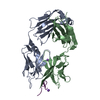
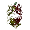
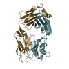
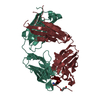
| Entry | Database: PDB / ID: 1acy | ||||||
|---|---|---|---|---|---|---|---|
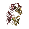
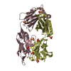
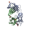
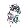
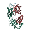
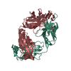
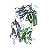
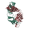
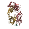
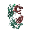
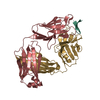
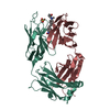
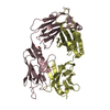
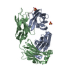
| Title | CRYSTAL STRUCTURE OF THE PRINCIPAL NEUTRALIZING SITE OF HIV-1 | ||||||
 Components Components |
| ||||||
 Keywords Keywords | COMPLEX(ANTIBODY/HIV-1 FRAGMENT) / COMPLEX(ANTIBODY-HIV-1 FRAGMENT) / COMPLEX(ANTIBODY-HIV-1 FRAGMENT) complex | ||||||
| Function / homology |  Function and homology information Function and homology informationInitial triggering of complement / Classical antibody-mediated complement activation / FCGR activation / Role of phospholipids in phagocytosis /  Regulation of Complement cascade / Regulation of actin dynamics for phagocytic cup formation / humoral immune response mediated by circulating immunoglobulin / phagocytosis, recognition / positive regulation of type IIa hypersensitivity / positive regulation of type I hypersensitivity ...Initial triggering of complement / Classical antibody-mediated complement activation / FCGR activation / Role of phospholipids in phagocytosis / Regulation of Complement cascade / Regulation of actin dynamics for phagocytic cup formation / humoral immune response mediated by circulating immunoglobulin / phagocytosis, recognition / positive regulation of type IIa hypersensitivity / positive regulation of type I hypersensitivity / antibody-dependent cellular cytotoxicity / Fc-gamma receptor I complex binding / phagocytosis, engulfment / immunoglobulin complex, circulating / IgG immunoglobulin complex / Dectin-2 family / immunoglobulin receptor binding / immunoglobulin mediated immune response / positive regulation of phagocytosis / complement activation, classical pathway / positive regulation of plasma membrane raft polarization / positive regulation of receptor clustering / antigen binding / positive regulation of establishment of T cell polarity / virus-mediated perturbation of host defense response / B cell differentiation / host cell endosome membrane / positive regulation of immune response / antibacterial humoral response / clathrin-dependent endocytosis of virus by host cell / viral protein processing / defense response to bacterium / fusion of virus membrane with host plasma membrane / external side of plasma membrane / fusion of virus membrane with host endosome membrane / viral envelope / virion attachment to host cell / host cell plasma membrane / virion membrane / structural molecule activity / extracellular space / membrane / identical protein binding / plasma membrane / cytoplasm Regulation of Complement cascade / Regulation of actin dynamics for phagocytic cup formation / humoral immune response mediated by circulating immunoglobulin / phagocytosis, recognition / positive regulation of type IIa hypersensitivity / positive regulation of type I hypersensitivity ...Initial triggering of complement / Classical antibody-mediated complement activation / FCGR activation / Role of phospholipids in phagocytosis / Regulation of Complement cascade / Regulation of actin dynamics for phagocytic cup formation / humoral immune response mediated by circulating immunoglobulin / phagocytosis, recognition / positive regulation of type IIa hypersensitivity / positive regulation of type I hypersensitivity / antibody-dependent cellular cytotoxicity / Fc-gamma receptor I complex binding / phagocytosis, engulfment / immunoglobulin complex, circulating / IgG immunoglobulin complex / Dectin-2 family / immunoglobulin receptor binding / immunoglobulin mediated immune response / positive regulation of phagocytosis / complement activation, classical pathway / positive regulation of plasma membrane raft polarization / positive regulation of receptor clustering / antigen binding / positive regulation of establishment of T cell polarity / virus-mediated perturbation of host defense response / B cell differentiation / host cell endosome membrane / positive regulation of immune response / antibacterial humoral response / clathrin-dependent endocytosis of virus by host cell / viral protein processing / defense response to bacterium / fusion of virus membrane with host plasma membrane / external side of plasma membrane / fusion of virus membrane with host endosome membrane / viral envelope / virion attachment to host cell / host cell plasma membrane / virion membrane / structural molecule activity / extracellular space / membrane / identical protein binding / plasma membrane / cytoplasmSimilarity search - Function | ||||||
| Biological species |  Mus musculus (house mouse) Mus musculus (house mouse) HIV-1 M:B_MN (virus) HIV-1 M:B_MN (virus) | ||||||
| Method | X-RAY DIFFRACTION / Resolution: 3 Å | ||||||
 Authors Authors | Ghiara, J.B. / Wilson, I.A. | ||||||
 Citation Citation | Journal: Science / Year: 1994 Title: Crystal structure of the principal neutralization site of HIV-1. Authors: Ghiara, J.B. / Stura, E.A. / Stanfield, R.L. / Profy, A.T. / Wilson, I.A. | ||||||
| History |
|
- Structure visualization
| Structure viewer | Molecule: MolmilJmol/JSmol |
|---|
- Downloads & links
-Download
| PDBx/mmCIF format | 1acy.cif.gz | 93.8 KB | Display | PDBx/mmCIF format |
|---|---|---|---|---|
| PDB format | pdb1acy.ent.gz | 76.2 KB | Display | PDB format |
| PDBx/mmJSON format | 1acy.json.gz | Tree view | PDBx/mmJSON format | |
| Others |  Other downloads Other downloads |
-Validation report
| Arichive directory | https://data.pdbj.org/pub/pdb/validation_reports/ac/1acyftp://data.pdbj.org/pub/pdb/validation_reports/ac/1acy | HTTPS FTP |
|---|
-Related structure data
| Similar structure data |
|---|


-Links
 PDBj
PDBj


- Assembly
| Deposited unit | 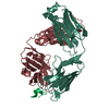
| ||||||||
|---|---|---|---|---|---|---|---|---|---|
| 1 |
| ||||||||
| Unit cell |
| ||||||||
| Atom site foot note | 1: CIS PROLINE - PRO L 8 / 2: CIS PROLINE - PRO L 77 / 3: CIS PROLINE - PRO L 95 / 4: CIS PROLINE - PRO L 141 / 5: CIS PROLINE - PRO H 149 / 6: CIS PROLINE - PRO H 151 / 7: CIS PROLINE - PRO H 200 |
-Components
| #1: Antibody | Mass: 23675.107 Da / Num. of mol.: 1 Source method: isolated from a genetically manipulated source Source: (gene. exp.) Mus musculus (house mouse) |
|---|---|
| #2: Antibody | Mass: 24208.383 Da / Num. of mol.: 1 Source method: isolated from a genetically manipulated source Source: (gene. exp.) Mus musculus (house mouse) / References: UniProt: P01869 |
| #3: Protein/peptide | Mass: 2814.339 Da / Num. of mol.: 1 / Fragment: FRAGMENT (RESIDUES 308 - 332) Source method: isolated from a genetically manipulated source Source: (gene. exp.) HIV-1 M:B_MN (virus) / Genus: Lentivirus / Species: Human immunodeficiency virus 1Subtypes of HIV / References: UniProt: P05877 |
| Compound details | THE PEPTIDE USED WAS A SYNTHETIC HOMOLOGUE OF RESIDUES 308 - 332 OF HIV-1 GP120 (MN STRAIN), WITH ...THE PEPTIDE USED WAS A SYNTHETIC HOMOLOGUE OF RESIDUES 308 - 332 OF HIV-1 GP120 (MN STRAIN), WITH AN ADDITIONAL |
| Sequence details | THE FAB FRAGMENT IS NUMBERED ACCORDING TO THE CONVENTION OF E. KABAT (E.A. KABAT, T.T. WU, M. REID- ...THE FAB FRAGMENT IS NUMBERED ACCORDING TO THE CONVENTION |
-Experimental details
-Experiment
| Experiment | Method: X-RAY DIFFRACTION |
|---|
- Sample preparation
| Crystal | Density Matthews: 4.15 Å3/Da / Density % sol: 70.36 % | ||||||||||||||||||||||||
|---|---|---|---|---|---|---|---|---|---|---|---|---|---|---|---|---|---|---|---|---|---|---|---|---|---|
| Crystal grow | *PLUS Temperature: 22 ℃ / pH: 6.75 / Method: vapor diffusion, sitting drop | ||||||||||||||||||||||||
| Components of the solutions | *PLUS
|
-Data collection
| Radiation | Scattering type: x-ray |
|---|---|
| Radiation wavelength | Relative weight: 1 |
| Reflection | *PLUS Highest resolution: 3 Å / Num. obs: 16265 / % possible obs: 94 % / Num. measured all: 79118 / Rmerge(I) obs: 0.116 |
- Processing
| Software |
| ||||||||||||||||||||||||||||||||||||||||||||||||||||||||||||
|---|---|---|---|---|---|---|---|---|---|---|---|---|---|---|---|---|---|---|---|---|---|---|---|---|---|---|---|---|---|---|---|---|---|---|---|---|---|---|---|---|---|---|---|---|---|---|---|---|---|---|---|---|---|---|---|---|---|---|---|---|---|
| Refinement | Resolution: 3→12 Å / σ(F): 0 /
| ||||||||||||||||||||||||||||||||||||||||||||||||||||||||||||
| Refinement step | Cycle: LAST / Resolution: 3→12 Å
| ||||||||||||||||||||||||||||||||||||||||||||||||||||||||||||
| Refine LS restraints |
| ||||||||||||||||||||||||||||||||||||||||||||||||||||||||||||
| Software | *PLUS Name: X-PLOR / Classification: refinement | ||||||||||||||||||||||||||||||||||||||||||||||||||||||||||||
| Refinement | *PLUS Num. reflection all: 15181 / Rfactor all: 0.21 / Rfactor Rwork: 0.21 | ||||||||||||||||||||||||||||||||||||||||||||||||||||||||||||
| Solvent computation | *PLUS | ||||||||||||||||||||||||||||||||||||||||||||||||||||||||||||
| Displacement parameters | *PLUS | ||||||||||||||||||||||||||||||||||||||||||||||||||||||||||||
| Refine LS restraints | *PLUS Type: x_angle_d / Dev ideal: 3.8 |
