Movie
Movie Controller

[English] 日本語
 Yorodumi
- EMDB-42730: Constituent EM map: Focused refinement TaF+TM+CTD of the Structur... -
+ Open data

- Basic information
| Entry |  | |||||||||
|---|---|---|---|---|---|---|---|---|---|---|
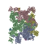
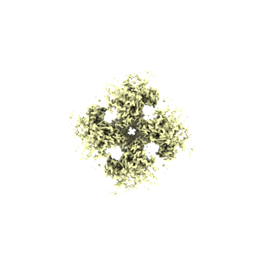
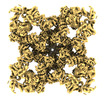
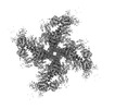
| Title | Constituent EM map: Focused refinement TaF+TM+CTD of the Structure of PKA phosphorylated human RyR2-R420W in the primed state in the presence of calcium | |||||||||
 Map data Map data | Constituent EM map: Focused refinement TaF TM CTD of the Structure of PKA phosphorylated human RyR2-R420W in the primed state in the presence of calcium | |||||||||
 Sample Sample |
| |||||||||
 Keywords Keywords |  calcium channel / MEMBRANE PROTEIN calcium channel / MEMBRANE PROTEIN | |||||||||
| Biological species |  Homo sapiens (human) Homo sapiens (human) | |||||||||
| Method | single particle reconstruction / cryo EM / Resolution: 3.21 Å | |||||||||
 Authors Authors | Miotto MC / Marks AR | |||||||||
| Funding support |  United States, 1 items United States, 1 items
| |||||||||
 Citation Citation | Journal: To Be Published Title: Structural basis for ryanodine receptor type 2 leak in heart failure and arrhythmogenic disorders Authors: Miotto MC | |||||||||
| History |
|
- Structure visualization
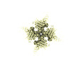
| Supplemental images |
|---|
- Downloads & links
-EMDB archive
| Map data | emd_42730.map.gz | 253.5 MB |  EMDB map data format EMDB map data format | |
|---|---|---|---|---|
| Header (meta data) | emd-42730-v30.xmlemd-42730.xml | 16.2 KB 16.2 KB | Display Display | EMDB header |
| Images |  emd_42730.png emd_42730.png | 45.2 KB | ||
| Filedesc metadata | emd-42730.cif.gz | 4.4 KB | ||
| Others | emd_42730_half_map_1.map.gzemd_42730_half_map_2.map.gz | 473.9 MB 473.9 MB | ||
| Archive directory |  http://ftp.pdbj.org/pub/emdb/structures/EMD-42730ftp://ftp.pdbj.org/pub/emdb/structures/EMD-42730 http://ftp.pdbj.org/pub/emdb/structures/EMD-42730ftp://ftp.pdbj.org/pub/emdb/structures/EMD-42730 | HTTPS FTP |
-Related structure data
| Related structure data |  8uq2C  8uq3C  8uq4C  8uq5C  8uxcC  8uxeC  8uxfC  8uxgC  8uxhC  8uxiC  8uxlC  8uxmC C: citing same article ( |
|---|


-Links
| EMDB pages | EMDB (EBI/PDBe) / EMDataResource |
|---|
-Map
| File | Download / File: emd_42730.map.gz / Format: CCP4 / Size: 512 MB / Type: IMAGE STORED AS FLOATING POINT NUMBER (4 BYTES) | ||||||||||||||||||||
|---|---|---|---|---|---|---|---|---|---|---|---|---|---|---|---|---|---|---|---|---|---|
| Annotation | Constituent EM map: Focused refinement TaF TM CTD of the Structure of PKA phosphorylated human RyR2-R420W in the primed state in the presence of calcium | ||||||||||||||||||||
| Voxel size | X=Y=Z: 0.83 Å | ||||||||||||||||||||
| Density |
| ||||||||||||||||||||
| Symmetry | Space group: 1 | ||||||||||||||||||||
| Details | EMDB XML:
|
-Supplemental data
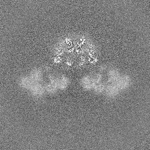
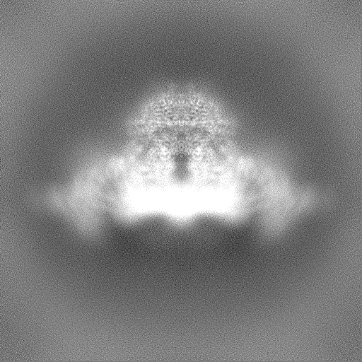
-Half map: #1
| File | emd_42730_half_map_1.map | ||||||||||||
|---|---|---|---|---|---|---|---|---|---|---|---|---|---|
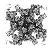
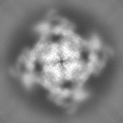
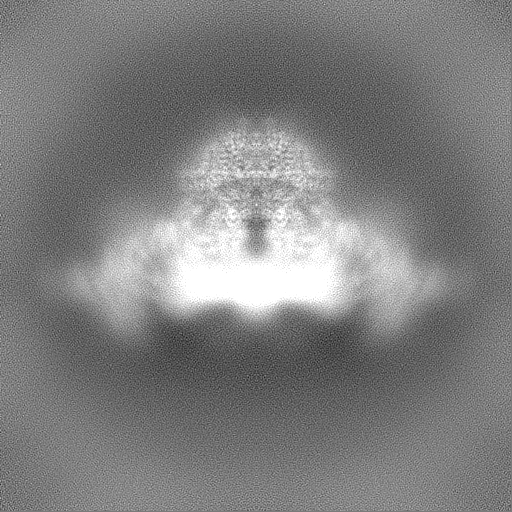
| Projections & Slices |
| ||||||||||||
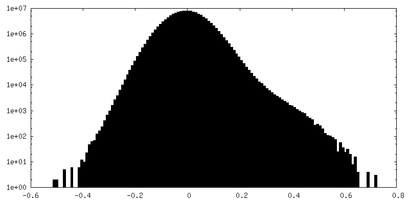
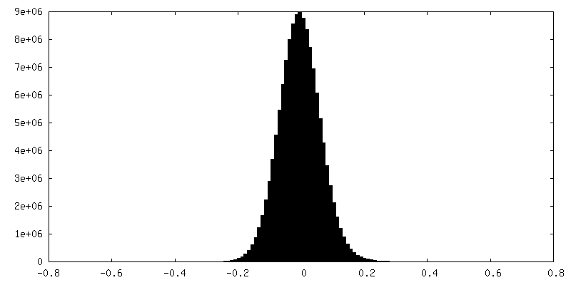
| Density Histograms |
 Z
Z Y
Y X
X


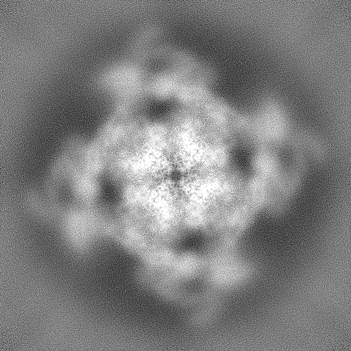
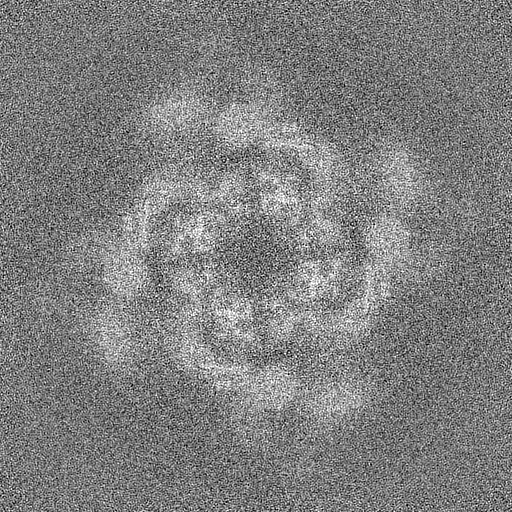
-Half map: #2
| File | emd_42730_half_map_2.map | ||||||||||||
|---|---|---|---|---|---|---|---|---|---|---|---|---|---|
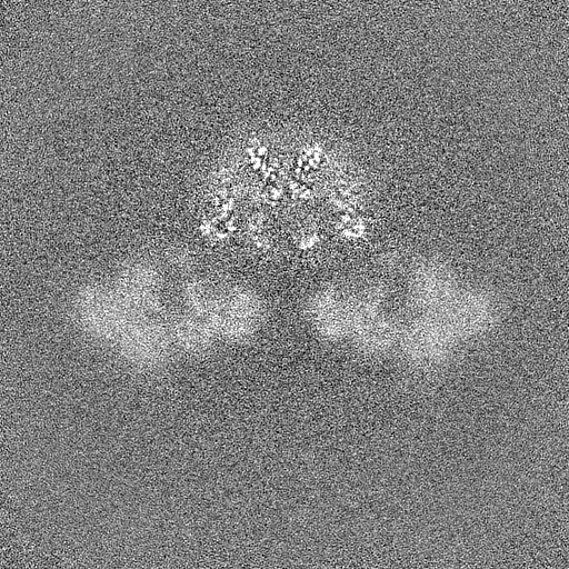
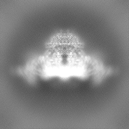
| Projections & Slices |
| ||||||||||||
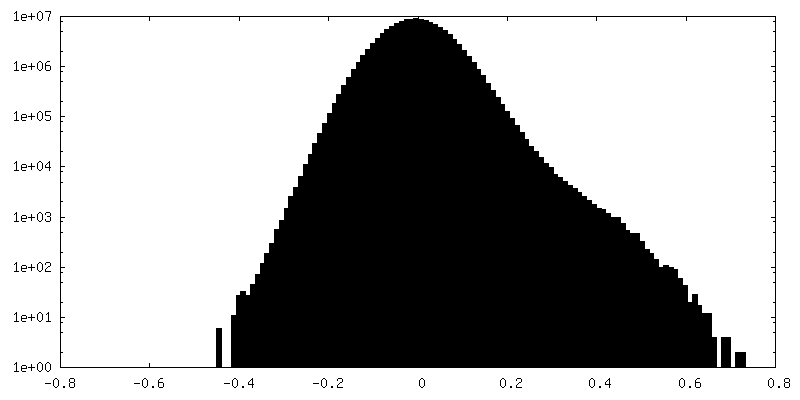
| Density Histograms |


- Sample components
-Entire : Complex of RyR2-R420W and Calstabin-2
| Entire | Name: Complex of RyR2-R420W and Calstabin-2 |
|---|---|
| Components |
|
-Supramolecule #1: Complex of RyR2-R420W and Calstabin-2
| Supramolecule | Name: Complex of RyR2-R420W and Calstabin-2 / type: complex / ID: 1 / Parent: 0 / Macromolecule list: #1-#2 |
|---|
-Supramolecule #2: Ryanodine Receptor 2
| Supramolecule | Name: Ryanodine Receptor 2 / type: complex / ID: 2 / Parent: 1 / Macromolecule list: #1 |
|---|---|
| Source (natural) | Organism: Homo sapiens (human) |
-Supramolecule #3: Peptidyl- cis-trans isomerase FKBP1B
| Supramolecule | Name: Peptidyl- cis-trans isomerase FKBP1B / type: complex / ID: 3 / Parent: 1 / Macromolecule list: #2 |
|---|---|
| Source (natural) | Organism: Homo sapiens (human) |
-Experimental details
-Structure determination
| Method | cryo EM |
|---|---|
 Processing Processing | single particle reconstruction |
| Aggregation state | particle |
-Sample preparation
| Concentration | 2.5 mg/mL | |||||||||||||||||||||||||||
|---|---|---|---|---|---|---|---|---|---|---|---|---|---|---|---|---|---|---|---|---|---|---|---|---|---|---|---|---|
| Buffer | pH: 7.4 Component:
| |||||||||||||||||||||||||||
| Vitrification | Cryogen name: ETHANE |
- Electron microscopy
| Microscope | FEI TITAN KRIOS |
|---|---|
| Electron beam | Acceleration voltage: 300 kV / Electron source: FIELD EMISSION GUN |
| Electron optics | C2 aperture diameter: 100.0 µm / Illumination mode: FLOOD BEAM / Imaging mode: BRIGHT FIELDBright-field microscopy / Cs: 2.7 mm / Nominal defocus max: 1.2 µm / Nominal defocus min: 0.5 µm |
| Specialist optics | Energy filter - Name: GIF Bioquantum / Energy filter - Slit width: 20 eV |
| Sample stage | Specimen holder model: FEI TITAN KRIOS AUTOGRID HOLDER / Cooling holder cryogen: NITROGEN |
| Temperature | Min: 80.0 K / Max: 100.0 K |
| Image recording | Film or detector model: GATAN K3 BIOQUANTUM (6k x 4k) / Digitization - Dimensions - Width: 5760 pixel / Digitization - Dimensions - Height: 4092 pixel / Average electron dose: 58.0 e/Å2 |
| Experimental equipment |  Model: Titan Krios / Image courtesy: FEI Company |
-Image processing
| Startup model | Type of model: INSILICO MODEL / In silico model: CryoSPARC ab initio |
|---|---|
| Initial angle assignment | Type: MAXIMUM LIKELIHOOD / Software - Name: cryoSPARC |
| Final angle assignment | Type: MAXIMUM LIKELIHOOD |
| Final reconstruction | Resolution.type: BY AUTHOR / Resolution: 3.21 Å / Resolution method: FSC 0.143 CUT-OFF / Software - Name: cryoSPARC / Number images used: 223544 |
-Atomic model buiding 1
| Initial model | PDB ID: Chain - Source name: PDB / Chain - Initial model type: experimental model |
|---|