Movie
Movie Controller
 Structure viewers
 About Yorodumi Papers


+Search query
-Structure paper
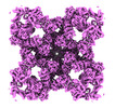
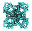
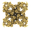
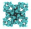
| Title | Structural basis for ryanodine receptor type 2 leak in heart failure and arrhythmogenic disorders |
|---|---|
| Journal, issue, pages | To Be Published |
| Publish date | Oct 23, 2023 (structure data deposition date) |
 Authors Authors | Miotto MC |
 External links External links | Search PubMed |
| Methods | EM (single particle) |
| Resolution | 2.79 - 5.65 Å |
| Structure data | EMDB-42458, PDB-8uq2: EMDB-42459, PDB-8uq3: EMDB-42460, PDB-8uq4: EMDB-42461, PDB-8uq5:  EMDB-42505: Raw consensus map of the Structure of human RyR2-S2808D in the subprimed state  EMDB-42581: Constituent EM map: Focused refinement TaF+TM+CTD of the Structure of human RyR2-S2808D in the subprimed state  EMDB-42582: Constituent EM map: Focused refinement Jsol+CSol of the Structure of human RyR2-S2808D in the subprimed state  EMDB-42583: Constituent EM map: Focused refinement NTD+SPRY+Calstabin-2 of the Structure of human RyR2-S2808D in the subprimed state  EMDB-42584: Constituent EM map: Focused refinement RY3&4 of the Structure of human RyR2-S2808D in the subprimed state  EMDB-42585: Constituent EM map: Focused refinement RY1&2 of the Structure of human RyR2-S2808D in the subprimed state  EMDB-42586: Constituent EM map: Focused refinement BSol2 of the Structure of human RyR2-S2808D in the subprimed state  EMDB-42703: Raw consensus map of the Structure of PKA phosphorylated human RyR2-R420Q in the closed state in the presence of ARM210  EMDB-42704: Constituent EM map: Focused refinement TaF+TM+CTD of the Structure of PKA phosphorylated human RyR2-R420Q in the closed state in the presence of ARM210  EMDB-42705: Constituent EM map: Focused refinement Jsol+CSol of the Structure of PKA phosphorylated human RyR2-R420Q in the closed state in the presence of ARM210  EMDB-42706: Constituent EM map: Focused refinement NTD+SPRY+Calstabin-2 of the Structure of PKA phosphorylated human RyR2-R420Q in the closed state in the presence of ARM210  EMDB-42707: Constituent EM map: Focused refinement BSol of the Structure of PKA phosphorylated human RyR2-R420Q in the closed state in the presence of ARM210  EMDB-42708: Constituent EM map: Focused refinement RY1&2 of the Structure of PKA phosphorylated human RyR2-R420Q in the closed state in the presence of ARM210  EMDB-42709: Constituent EM map: Focused refinement RY3&4 of the Structure of PKA phosphorylated human RyR2-R420Q in the closed state in the presence of ARM210  EMDB-42710: Constituent EM map: Focused refinement BSol2 of the Structure of PKA phosphorylated human RyR2-R420Q in the closed state in the presence of ARM210  EMDB-42711: Raw consensus map of the Structure of PKA phosphorylated human RyR2-R420W in the primed state  EMDB-42712: Constituent EM map: Focused refinement TaF+TM+CTD of the Structure of PKA phosphorylated human RyR2-R420W in the primed state  EMDB-42713: Constituent EM map: Focused refinement Jsol+CSol of the Structure of PKA phosphorylated human RyR2-R420W in the primed state  EMDB-42715: Constituent EM map: Focused refinement NTD+SPRY+Calstabin-2 of the Structure of PKA phosphorylated human RyR2-R420W in the primed state  EMDB-42716: Constituent EM map: Focused refinement RY1&2 of the Structure of PKA phosphorylated human RyR2-R420W in the primed state  EMDB-42717: Constituent EM map: Focused refinement RY3&4 of the Structure of PKA phosphorylated human RyR2-R420W in the primed state  EMDB-42718: Constituent EM map: Focused refinement BSol2 of the Structure of PKA phosphorylated human RyR2-R420W in the primed state  EMDB-42722: Raw consensus map of the Structure of PKA phosphorylated human RyR2-R420W in the closed state in the presence of ARM210  EMDB-42723: Constituent EM map: Focused refinement TaF+TM+CTD of the Structure of PKA phosphorylated human RyR2-R420W in the closed state in the presence of ARM210  EMDB-42724: Constituent EM map: Focused refinement Jsol+CSol of the Structure of PKA phosphorylated human RyR2-R420W in the closed state in the presence of ARM210  EMDB-42725: Constituent EM map: Focused refinement NTD+SPRY+Calstabin-2 of the Structure of PKA phosphorylated human RyR2-R420W in the closed state in the presence of ARM210  EMDB-42726: Constituent EM map: Focused refinement RY1&2 of the Structure of PKA phosphorylated human RyR2-R420W in the closed state in the presence of ARM210  EMDB-42727: Constituent EM map: Focused refinement RY3&4 of the Structure of PKA phosphorylated human RyR2-R420W in the closed state in the presence of ARM210  EMDB-42728: Constituent EM map: Focused refinement BSol2 of the Structure of PKA phosphorylated human RyR2-R420W in the closed state in the presence of ARM210  EMDB-42729: Raw consensus map of the Structure of PKA phosphorylated human RyR2-R420W in the primed state in the presence of calcium  EMDB-42730: Constituent EM map: Focused refinement TaF+TM+CTD of the Structure of PKA phosphorylated human RyR2-R420W in the primed state in the presence of calcium  EMDB-42731: Constituent EM map: Focused refinement Jsol+CSol of the Structure of PKA phosphorylated human RyR2-R420W in the primed state in the presence of calcium  EMDB-42732: Constituent EM map: Focused refinement NTD+SPRY+Calstabin-2 of the Structure of PKA phosphorylated human RyR2-R420W in the primed state in the presence of calcium  EMDB-42733: Constituent EM map: Focused refinement RY1&2 of the Structure of PKA phosphorylated human RyR2-R420W in the primed state in the presence of calcium 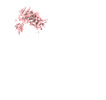 EMDB-42734: Constituent EM map: Focused refinement RY3&4 of the Structure of PKA phosphorylated human RyR2-R420W in the primed state in the presence of calcium  EMDB-42735: Constituent EM map: Focused refinement BSol2 of the Structure of PKA phosphorylated human RyR2-R420W in the primed state in the presence of calcium 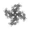 EMDB-42736: Raw consensus map of the Structure of PKA phosphorylated human RyR2-R420W in the open state in the presence of calcium 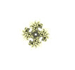 EMDB-42737: Constituent EM map: Focused refinement TaF+TM+CTD of the Structure of PKA phosphorylated human RyR2-R420W in the open state in the presence of calcium 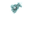 EMDB-42738: Constituent EM map: Focused refinement JSol+CSol of the Structure of PKA phosphorylated human RyR2-R420W in the open state in the presence of calcium  EMDB-42739: Constituent EM map: Focused refinement NTD+SPRY+Calstabin-2 of the Structure of PKA phosphorylated human RyR2-R420W in the open state in the presence of calcium  EMDB-42740: Constituent EM map: Focused refinement RY1&2 of the Structure of PKA phosphorylated human RyR2-R420W in the open state in the presence of calcium  EMDB-42741: Constituent EM map: Focused refinement RY3&4 of the Structure of PKA phosphorylated human RyR2-R420W in the open state in the presence of calcium  EMDB-42742: Constituent EM map: Focused refinement BSol2 of the Structure of PKA phosphorylated human RyR2-R420W in the open state in the presence of calcium EMDB-42759, PDB-8uxc: EMDB-42761, PDB-8uxe: EMDB-42762, PDB-8uxf: EMDB-42763, PDB-8uxg: EMDB-42764, PDB-8uxh: EMDB-42765, PDB-8uxi: EMDB-42768, PDB-8uxl: EMDB-42769, PDB-8uxm: |
| Chemicals |  ChemComp-ZN:  ChemComp-ATP: 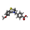 ChemComp-KVR:  ChemComp-CA: |
| Source |
|
 Keywords Keywords |  MEMBRANE PROTEIN / calcium channel / TRANSPORT PROTEIN MEMBRANE PROTEIN / calcium channel / TRANSPORT PROTEIN |