 Movie
Movie Controller

[English] 日本語
 Yorodumi
- EMDB-40204: CryoEM map of the locally refined interfaces-1,2,3 of soluble OPA... -
+ Open data

- Basic information
| Entry |  | |||||||||
|---|---|---|---|---|---|---|---|---|---|---|
| Title | CryoEM map of the locally refined interfaces-1,2,3 of soluble OPA1 from the apo bound helical assembly on a lipid membrane | |||||||||
 Map data Map data | CryoEM map of the locally refined interfaces-1,2,3 of soluble OPA1 from the apo bound helical assembly on a lipid membrane | |||||||||
 Sample Sample |
| |||||||||
 Keywords Keywords |  GTPase / Dynamin-family protein / mitochondrial fusion protein / mitochondria / Optic Atrophy / LIPID BINDING PROTEIN GTPase / Dynamin-family protein / mitochondrial fusion protein / mitochondria / Optic Atrophy / LIPID BINDING PROTEIN | |||||||||
| Function / homology |  Function and homology informationRegulation of Apoptosis / membrane tubulation / inner mitochondrial membrane organization / dynamin GTPase / cardiolipin binding / mitochondrial genome maintenance / phosphatidic acid binding / mitochondrial fission / GTP metabolic process / mitochondrial fusion ...Regulation of Apoptosis / membrane tubulation / inner mitochondrial membrane organization / dynamin GTPase / cardiolipin binding / mitochondrial genome maintenance / phosphatidic acid binding / mitochondrial fission / GTP metabolic process / mitochondrial fusion / axonal transport of mitochondrion / negative regulation of release of cytochrome c from mitochondria / mitochondrial crista / negative regulation of endoplasmic reticulum stress-induced intrinsic apoptotic signaling pathway / axon cytoplasm / visual perception / mitochondrion organization / neural tube closure / mitochondrial membrane / mitochondrial intermembrane space / cellular senescence / protein complex oligomerization / microtubule binding / mitochondrial inner membrane / microtubule / mitochondrial outer membrane / GTPase activity / dendrite / apoptotic process / GTP binding / negative regulation of apoptotic process / magnesium ion binding / mitochondrion / nucleoplasm / membrane / cytosol / cytoplasm Function and homology informationRegulation of Apoptosis / membrane tubulation / inner mitochondrial membrane organization / dynamin GTPase / cardiolipin binding / mitochondrial genome maintenance / phosphatidic acid binding / mitochondrial fission / GTP metabolic process / mitochondrial fusion ...Regulation of Apoptosis / membrane tubulation / inner mitochondrial membrane organization / dynamin GTPase / cardiolipin binding / mitochondrial genome maintenance / phosphatidic acid binding / mitochondrial fission / GTP metabolic process / mitochondrial fusion / axonal transport of mitochondrion / negative regulation of release of cytochrome c from mitochondria / mitochondrial crista / negative regulation of endoplasmic reticulum stress-induced intrinsic apoptotic signaling pathway / axon cytoplasm / visual perception / mitochondrion organization / neural tube closure / mitochondrial membrane / mitochondrial intermembrane space / cellular senescence / protein complex oligomerization / microtubule binding / mitochondrial inner membrane / microtubule / mitochondrial outer membrane / GTPase activity / dendrite / apoptotic process / GTP binding / negative regulation of apoptotic process / magnesium ion binding / mitochondrion / nucleoplasm / membrane / cytosol / cytoplasmSimilarity search - Function | |||||||||
| Biological species |  Homo sapiens (human) Homo sapiens (human) | |||||||||
| Method | helical reconstruction / cryo EM / Resolution: 5.51 Å | |||||||||
 Authors Authors | Nyenhuis SB / Wu X / Strub MP / Yim YI / Stanton AE / Baena V / Syed ZA / Canagarajah B / Hammer JA / Hinshaw JE | |||||||||
| Funding support |  United States, 1 items United States, 1 items
| |||||||||
 Citation Citation | Journal: Nature / Year: 2023 Title: OPA1 helical structures give perspective to mitochondrial dysfunction. Authors: Sarah B Nyenhuis / Xufeng Wu / Marie-Paule Strub / Yang-In Yim / Abigail E Stanton / Valentina Baena / Zulfeqhar A Syed / Bertram Canagarajah / John A Hammer / Jenny E Hinshaw / Abstract: Dominant optic atrophy is one of the leading causes of childhood blindness. Around 60-80% of cases are caused by mutations of the gene that encodes optic atrophy protein 1 (OPA1), a protein that has ...Dominant optic atrophy is one of the leading causes of childhood blindness. Around 60-80% of cases are caused by mutations of the gene that encodes optic atrophy protein 1 (OPA1), a protein that has a key role in inner mitochondrial membrane fusion and remodelling of cristae and is crucial for the dynamic organization and regulation of mitochondria. Mutations in OPA1 result in the dysregulation of the GTPase-mediated fusion process of the mitochondrial inner and outer membranes. Here we used cryo-electron microscopy methods to solve helical structures of OPA1 assembled on lipid membrane tubes, in the presence and absence of nucleotide. These helical assemblies organize into densely packed protein rungs with minimal inter-rung connectivity, and exhibit nucleotide-dependent dimerization of the GTPase domains-a hallmark of the dynamin superfamily of proteins. OPA1 also contains several unique secondary structures in the paddle domain that strengthen its membrane association, including membrane-inserting helices. The structural features identified in this study shed light on the effects of pathogenic point mutations on protein folding, inter-protein assembly and membrane interactions. Furthermore, mutations that disrupt the assembly interfaces and membrane binding of OPA1 cause mitochondrial fragmentation in cell-based assays, providing evidence of the biological relevance of these interactions. | |||||||||
| History |
|
- Structure visualization
| Supplemental images |
|---|

- Downloads & links
-EMDB archive
| Map data | emd_40204.map.gz | 208.3 MB | EMDB map data format | |
|---|---|---|---|---|
| Header (meta data) | emd-40204-v30.xmlemd-40204.xml | 20.5 KB 20.5 KB | Display Display | EMDB header |
| FSC (resolution estimation) | emd_40204_fsc.xml | 16 KB | Display | FSC data file |
| Images | 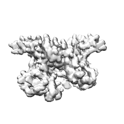 emd_40204.png emd_40204.png | 62.6 KB | ||
| Masks | emd_40204_msk_1.map | 421.9 MB | Mask map | |
| Others | emd_40204_additional_1.map.gzemd_40204_additional_2.map.gzemd_40204_half_map_1.map.gzemd_40204_half_map_2.map.gz | 352.2 MB 398.3 MB 391 MB 391 MB | ||
| Archive directory |  http://ftp.pdbj.org/pub/emdb/structures/EMD-40204ftp://ftp.pdbj.org/pub/emdb/structures/EMD-40204 http://ftp.pdbj.org/pub/emdb/structures/EMD-40204ftp://ftp.pdbj.org/pub/emdb/structures/EMD-40204 | HTTPS FTP |
-Related structure data
| Related structure data |  8eewC  8ef7C  8effC  8efrC  8efsC  8eftC C: citing same article ( |
|---|---|
| Similar structure data |


-Links
| EMDB pages | EMDB (EBI/PDBe) / EMDataResource |
|---|
-Map
| File | Download / File: emd_40204.map.gz / Format: CCP4 / Size: 421.9 MB / Type: IMAGE STORED AS FLOATING POINT NUMBER (4 BYTES) | ||||||||||||||||||||
|---|---|---|---|---|---|---|---|---|---|---|---|---|---|---|---|---|---|---|---|---|---|
| Annotation | CryoEM map of the locally refined interfaces-1,2,3 of soluble OPA1 from the apo bound helical assembly on a lipid membrane | ||||||||||||||||||||
| Voxel size | X=Y=Z: 1.2518 Å | ||||||||||||||||||||
| Density |
| ||||||||||||||||||||
| Symmetry | Space group: 1 | ||||||||||||||||||||
| Details | EMDB XML:
|
-Supplemental data
-Mask #1
| File | emd_40204_msk_1.map | ||||||||||||
|---|---|---|---|---|---|---|---|---|---|---|---|---|---|
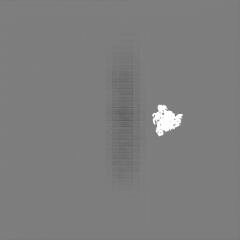
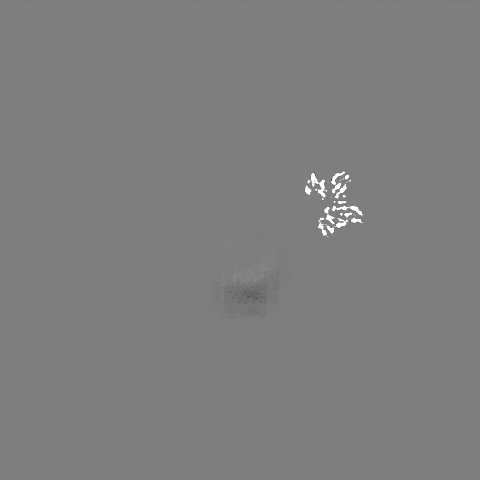
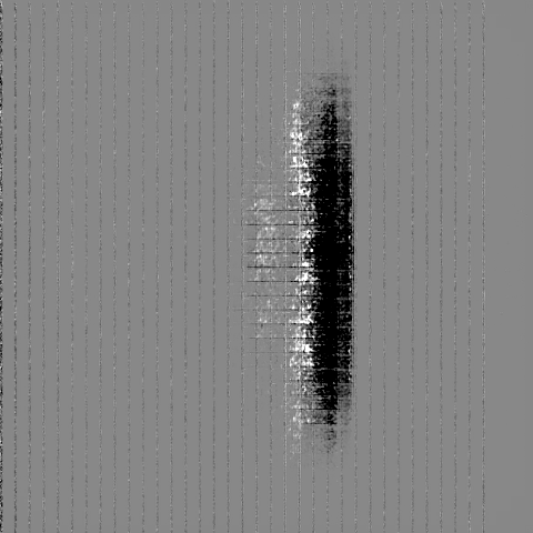
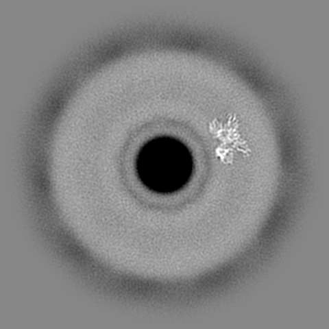
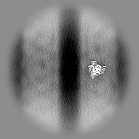
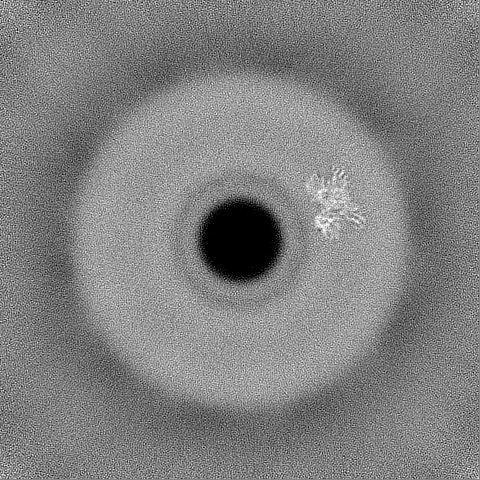
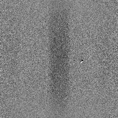
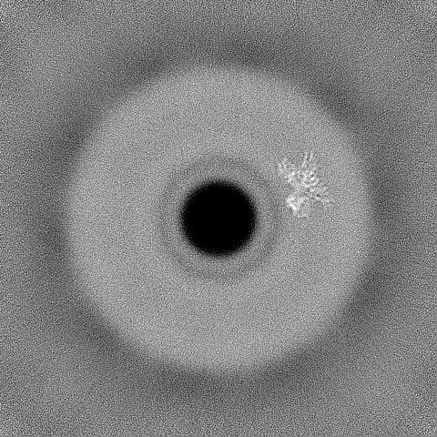
| Projections & Slices |
| ||||||||||||
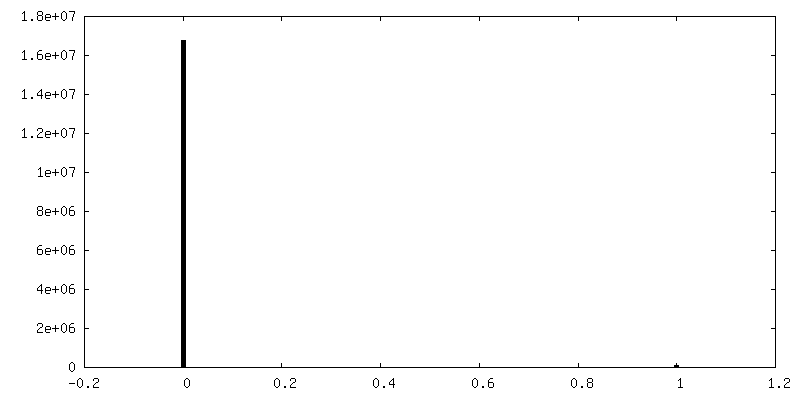
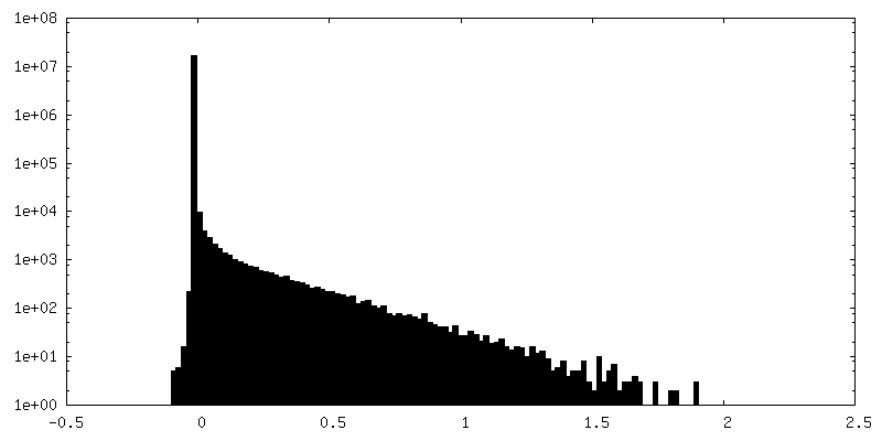
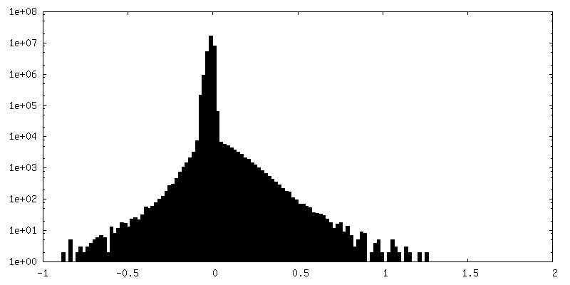
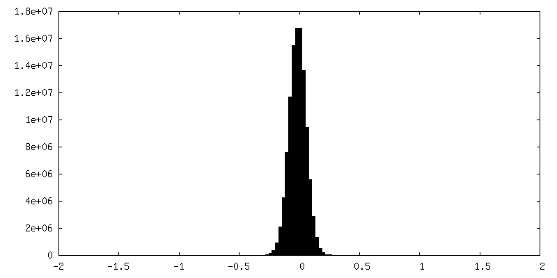
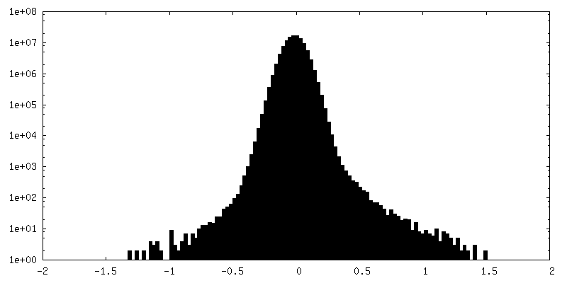
| Density Histograms |
 Z
Z Y
Y X
X


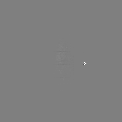
-Additional map: CryoEM sharpened map 1 of the locally refined...
| File | emd_40204_additional_1.map | ||||||||||||
|---|---|---|---|---|---|---|---|---|---|---|---|---|---|
| Annotation | CryoEM sharpened map 1 of the locally refined interfaces-1,2,3 of soluble OPA1 from the apo bound helical assembly on a lipid membrane | ||||||||||||
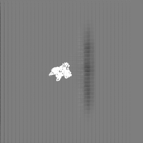
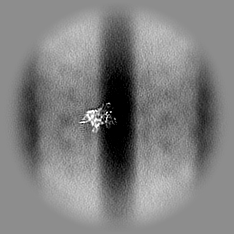
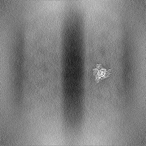
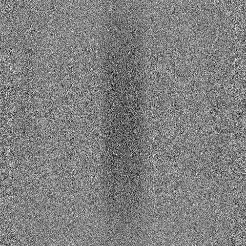
| Projections & Slices |
| ||||||||||||
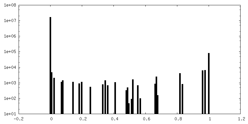
| Density Histograms |


-Additional map: CryoEM sharpened map 2 of the locally refined...
| File | emd_40204_additional_2.map | ||||||||||||
|---|---|---|---|---|---|---|---|---|---|---|---|---|---|
| Annotation | CryoEM sharpened map 2 of the locally refined interfaces-1,2,3 of soluble OPA1 from the apo bound helical assembly on a lipid membrane | ||||||||||||
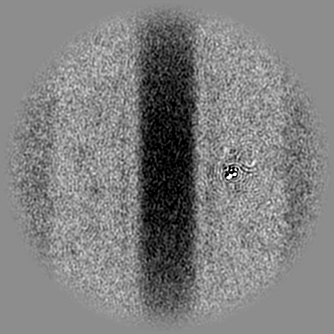
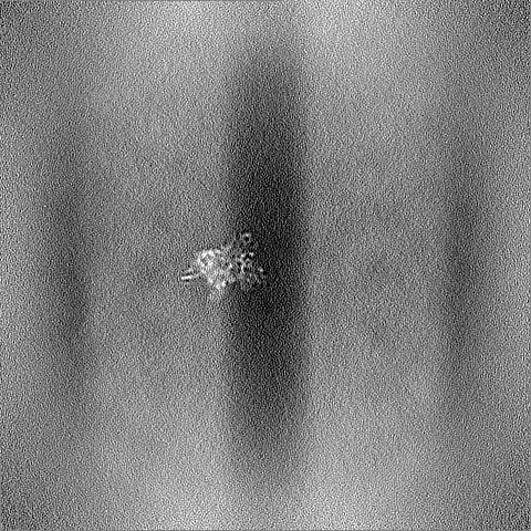
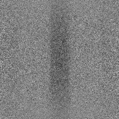
| Projections & Slices |
| ||||||||||||
| Density Histograms |


-Half map: CryoEM half map A of the locally refined...
| File | emd_40204_half_map_1.map | ||||||||||||
|---|---|---|---|---|---|---|---|---|---|---|---|---|---|
| Annotation | CryoEM half map A of the locally refined interfaces-1,2,3 of soluble OPA1 from the apo bound helical assembly on a lipid membrane | ||||||||||||
| Projections & Slices |
| ||||||||||||
| Density Histograms |


-Half map: CryoEM half map B of the locally refined...
| File | emd_40204_half_map_2.map | ||||||||||||
|---|---|---|---|---|---|---|---|---|---|---|---|---|---|
| Annotation | CryoEM half map B of the locally refined interfaces-1,2,3 of soluble OPA1 from the apo bound helical assembly on a lipid membrane | ||||||||||||
| Projections & Slices |
| ||||||||||||
| Density Histograms |


- Sample components
-Entire : CryoEM map of the locally refined soluble OPA1 Z-clip from the ap...
| Entire | Name: CryoEM map of the locally refined soluble OPA1 Z-clip from the apo helical assembly on a lipid membrane |
|---|---|
| Components |
|
-Supramolecule #1: CryoEM map of the locally refined soluble OPA1 Z-clip from the ap...
| Supramolecule | Name: CryoEM map of the locally refined soluble OPA1 Z-clip from the apo helical assembly on a lipid membrane type: complex / ID: 1 / Parent: 0 / Macromolecule list: all |
|---|---|
| Source (natural) | Organism: Homo sapiens (human) |
-Macromolecule #1: sOPA1 protein
| Macromolecule | Name: sOPA1 protein / type: protein_or_peptide / ID: 1 / Enantiomer: LEVO |
|---|---|
| Source (natural) | Organism: Homo sapiens (human) |
| Recombinant expression | Organism:  Escherichia coli BL21(DE3) (bacteria) Escherichia coli BL21(DE3) (bacteria) |
| Sequence | String: ATDRGSESDK HFRKVSDKEK IDQLQEELLH TQLKYQRILE RLEKENKELR KLVLQKDDKG IHHRKLKKSL IDMYSEVLD VLSDYDASYN TQDHLPRVVV VGDQSAGKTS VLEMIAQARI FPRGSGEMMT RSPVKVTLSE G PHHVALFK DSSREFDLTK EEDLAALRHE ...String: ATDRGSESDK HFRKVSDKEK IDQLQEELLH TQLKYQRILE RLEKENKELR KLVLQKDDKG IHHRKLKKSL IDMYSEVLD VLSDYDASYN TQDHLPRVVV VGDQSAGKTS VLEMIAQARI FPRGSGEMMT RSPVKVTLSE G PHHVALFK DSSREFDLTK EEDLAALRHE IELRMRKNVK EGCTVSPETI SLNVKGPGLQ RMVLVDLPGV IN TVTSGMA PDTKETIFSI SKAYMQNPNA IILCIQDGSV DAERSIVTDL VSQMDPHGRR TIFVLTKVDL AEK NVASPS RIQQIIEGKL FPMKALGYFA VVTGKGNSSE SIEAIREYEE EFFQNSKLLK TSMLKAHQVT TRNL SLAVS DCFWKMVRES VEQQADSFKA TRFNLETEWK NNYPRLRELD RNELFEKAKN EILDEVISLS QVTPK HWEE ILQQSLWERV STHVIENIYL PAAQTMNSGT FNTTVDIKLK QWTDKQLPNK AVEVAWETLQ EEFSRF MTE PKGKEHDDIF DKLKEAVKEE SIKRHKWNDF AEDSLRVIQH NALEDRSISD KQQWDAAIYF MEEALQA RL KDTENAIENM VGPDWKKRWL YWKNRTQEQC VHNETKNELE KMLKCNEEHP AYLASDEITT VRKNLESR G VEVDPSLIKD TWHQVYRRHF LKTALNHCNL CRRGFYYYQR HFVDSELECN DVVLFWRIQR MLAITANTL RQQLTNTEVR RLEKNVKEVL EDFAEDGEKK IKLLTGKRVQ LAEDLKKVRE IQEKLDAFIE ALHQEK UniProtKB: Dynamin-like 120 kDa protein, mitochondrial |
-Experimental details
-Structure determination
| Method | cryo EM |
|---|---|
 Processing Processing | helical reconstruction |
| Aggregation state | helical array |
-Sample preparation
| Buffer | pH: 7.2 |
|---|---|
| Vitrification | Cryogen name: ETHANE |
- Electron microscopy
| Microscope | TFS GLACIOS |
|---|---|
| Electron beam | Acceleration voltage: 200 kV / Electron source: FIELD EMISSION GUN |
| Electron optics | Illumination mode: FLOOD BEAM / Imaging mode: BRIGHT FIELDBright-field microscopy / Nominal defocus max: 2.4 µm / Nominal defocus min: 0.6 µm |
| Image recording | Film or detector model: GATAN K3 BIOQUANTUM (6k x 4k) / Average electron dose: 24.86 e/Å2 |
-Image processing
| Startup model | Type of model: PDB ENTRY PDB model - PDB ID: |
|---|---|
| Final angle assignment | Type: NOT APPLICABLE |
| Final reconstruction | Applied symmetry - Helical parameters - Δz: 14.07 Å Applied symmetry - Helical parameters - Δ&Phi: 37.25 ° Applied symmetry - Helical parameters - Axial symmetry: C1 (asymmetric) Resolution.type: BY AUTHOR / Resolution: 5.51 Å / Resolution method: FSC 0.143 CUT-OFF / Software - Name: cryoSPARC / Number images used: 352618 |
| FSC plot (resolution estimation) | 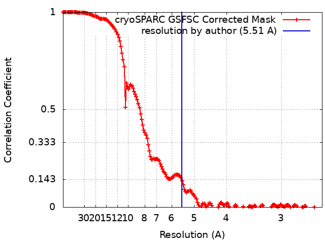 |

-Atomic model buiding 1
| Refinement | Space: REAL / Protocol: FLEXIBLE FIT |
|---|
