ムービー
ムービー コントローラー
 構造ビューア
 万見文献について


+検索条件
-Structure paper
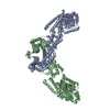
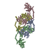
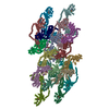
| タイトル | OPA1 helical structures give perspective to mitochondrial dysfunction. |
|---|---|
| ジャーナル・号・ページ | Nature, Vol. 620, Issue 7976, Page 1109-1116, Year 2023 |
| 掲載日 | 2023年8月23日 |
 著者 著者 | Sarah B Nyenhuis / Xufeng Wu / Marie-Paule Strub / Yang-In Yim / Abigail E Stanton / Valentina Baena / Zulfeqhar A Syed / Bertram Canagarajah / John A Hammer / Jenny E Hinshaw /  |
| PubMed 要旨 | Dominant optic atrophy is one of the leading causes of childhood blindness. Around 60-80% of cases are caused by mutations of the gene that encodes optic atrophy protein 1 (OPA1), a protein that has ...Dominant optic atrophy is one of the leading causes of childhood blindness. Around 60-80% of cases are caused by mutations of the gene that encodes optic atrophy protein 1 (OPA1), a protein that has a key role in inner mitochondrial membrane fusion and remodelling of cristae and is crucial for the dynamic organization and regulation of mitochondria. Mutations in OPA1 result in the dysregulation of the GTPase-mediated fusion process of the mitochondrial inner and outer membranes. Here we used cryo-electron microscopy methods to solve helical structures of OPA1 assembled on lipid membrane tubes, in the presence and absence of nucleotide. These helical assemblies organize into densely packed protein rungs with minimal inter-rung connectivity, and exhibit nucleotide-dependent dimerization of the GTPase domains-a hallmark of the dynamin superfamily of proteins. OPA1 also contains several unique secondary structures in the paddle domain that strengthen its membrane association, including membrane-inserting helices. The structural features identified in this study shed light on the effects of pathogenic point mutations on protein folding, inter-protein assembly and membrane interactions. Furthermore, mutations that disrupt the assembly interfaces and membrane binding of OPA1 cause mitochondrial fragmentation in cell-based assays, providing evidence of the biological relevance of these interactions. |
 リンク リンク | Nature / PubMed:37612506 |
| 手法 | EM (らせん対称) |
| 解像度 | 2.88 - 9.68 Å |
| 構造データ | EMDB-28063, PDB-8eew: EMDB-28074, PDB-8ef7:  EMDB-40192: CryoEM map of the locally refined soluble OPA1 Z-clip from the GDP-AlFx bound helical assembly on a lipid membrane  EMDB-40193: CryoEM map of the locally refined soluble OPA1 Z-clip from the apo helical assembly on a lipid membrane  EMDB-40197: CryoEM map of soluble OPA1 from the GDP-AlFx bound N-terminal helical assembly on a lipid membrane  EMDB-40198: CryoEM map of the locally refined interface-8 of soluble OPA1 from the GDP-AlFx bound N-terminal helical assembly on a lipid membrane  EMDB-40200: CryoEM map of the locally refined dimer of soluble OPA1 from the GDP-AlFx bound helical assembly on a lipid membrane  EMDB-40202: CryoEM map of the locally refined dimer of soluble OPA1 from the apo bound helical assembly on a lipid membrane  EMDB-40203: CryoEM map of the locally refined interfaces-1,2,3 of soluble OPA1 from the GDP-AlFx bound helical assembly on a lipid membrane  EMDB-40204: CryoEM map of the locally refined interfaces-1,2,3 of soluble OPA1 from the apo bound helical assembly on a lipid membrane  EMDB-40210: CryoEM map of the locally refined interface-4 of soluble OPA1 from the GDP-AlFx bound helical assembly on a lipid membrane  EMDB-40211: CryoEM map of the locally refined interface-4 of soluble OPA1 from the apo bound helical assembly on a lipid membrane  EMDB-40212: CryoEM map of the locally refined interface-7 of soluble OPA1 from the GDP-AlFx bound helical assembly on a lipid membrane  EMDB-40213: CryoEM map of the locally refined interface-7 of soluble OPA1 from the apo bound helical assembly on a lipid membrane  EMDB-40214: CryoEM map of the locally refined interfaces-5,6 of soluble OPA1 from the GDP-AlFx bound helical assembly on a lipid membrane  EMDB-40215: CryoEM map of the locally refined monomer-A of soluble OPA1 from the GDP-AlFx bound helical assembly on a lipid membrane  EMDB-40216: CryoEM map of the locally refined monomer-B of soluble OPA1 from the GDP-AlFx bound helical assembly on a lipid membrane  EMDB-40217: CryoEM map of the locally refined cardiolipin containing monolayer and soluble OPA1 paddles from the GDP-AlFx bound helical assembly on a lipid membrane |
| 化合物 |  ChemComp-GDP:  ChemComp-ALF:  ChemComp-MG:  ChemComp-K: |
| 由来 |
|
 キーワード キーワード | LIPID BINDING PROTEIN /  GTPase (GTPアーゼ) / Dynamin-family protein / mitochondrial fusion protein / mitochondria (ミトコンドリア) / Optic Atrophy GTPase (GTPアーゼ) / Dynamin-family protein / mitochondrial fusion protein / mitochondria (ミトコンドリア) / Optic Atrophy |