 Movie
Movie Controller

[English] 日本語
 Yorodumi
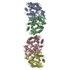
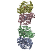
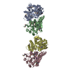
- EMDB-25835: CryoEM Structure of sFab COP-3 Complex with human claudin-4 and C... -
+ Open data

- Basic information
| Entry | Database: EMDB / ID: EMD-25835 | |||||||||
|---|---|---|---|---|---|---|---|---|---|---|
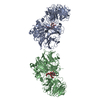
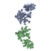
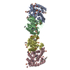
| Title | CryoEM Structure of sFab COP-3 Complex with human claudin-4 and Clostridium perfringens enterotoxin C-terminal domain | |||||||||
 Map data Map data | ||||||||||
 Sample Sample |
| |||||||||
 Keywords Keywords |  Claudin / cell adhesion / enterotoxin / Fab / antibody fragment / MEMBRANE PROTEIN Claudin / cell adhesion / enterotoxin / Fab / antibody fragment / MEMBRANE PROTEIN | |||||||||
| Function / homology |  Function and homology information Function and homology informationpositive regulation of metallopeptidase activity / calcium-independent cell-cell adhesion via plasma membrane cell-adhesion molecules / Tight junction interactions / apicolateral plasma membrane / bicellular tight junction assembly / regulation of cell morphogenesis / tight junction / chloride channel activity / positive regulation of wound healing / renal absorption ...positive regulation of metallopeptidase activity / calcium-independent cell-cell adhesion via plasma membrane cell-adhesion molecules / Tight junction interactions / apicolateral plasma membrane / bicellular tight junction assembly / regulation of cell morphogenesis / tight junction / chloride channel activity / positive regulation of wound healing / renal absorption / chloride channel complex / lateral plasma membrane / bicellular tight junction / establishment of skin barrier / basal plasma membrane / response to progesterone / female pregnancy / circadian rhythm / cell-cell junction / transmembrane signaling receptor activity / toxin activity / cell adhesion / positive regulation of cell migration / apical plasma membrane / structural molecule activity / extracellular region / identical protein binding / plasma membraneSimilarity search - Function | |||||||||
| Biological species |  Homo sapiens (human) / Homo sapiens (human) /   Clostridium perfringens (bacteria) Clostridium perfringens (bacteria) | |||||||||
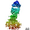
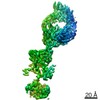
| Method | single particle reconstruction / cryo EM / Resolution: 5.0 Å | |||||||||
 Authors Authors | Vecchio AJ / Orlando BJ | |||||||||
| Funding support |  United States, 2 items United States, 2 items
| |||||||||
 Citation Citation | Journal: J Biol Chem / Year: 2022 Title: Development, structure, and mechanism of synthetic antibodies that target claudin and Clostridium perfringens enterotoxin complexes. Authors: Benjamin J Orlando / Pawel K Dominik / Sourav Roy / Chinemerem P Ogbu / Satchal K Erramilli / Anthony A Kossiakoff / Alex J Vecchio / Abstract: Strains of Clostridium perfringens produce a two-domain enterotoxin (CpE) that afflicts humans and domesticated animals, causing prevalent gastrointestinal illnesses. CpE's C-terminal domain (cCpE) ...Strains of Clostridium perfringens produce a two-domain enterotoxin (CpE) that afflicts humans and domesticated animals, causing prevalent gastrointestinal illnesses. CpE's C-terminal domain (cCpE) binds cell surface receptors, followed by a restructuring of its N-terminal domain to form a membrane-penetrating β-barrel pore, which is toxic to epithelial cells of the gut. The claudin family of membrane proteins are known receptors for CpE and also control the architecture and function of cell-cell contacts (tight junctions) that create barriers to intercellular molecular transport. CpE binding and assembly disables claudin barrier function and induces cytotoxicity via β-pore formation, disrupting gut homeostasis; however, a structural basis of this process and strategies to inhibit the claudin-CpE interactions that trigger it are both lacking. Here, we used a synthetic antigen-binding fragment (sFab) library to discover two sFabs that bind claudin-4 and cCpE complexes. We established these sFabs' mode of molecular recognition and binding properties and determined structures of each sFab bound to claudin-4-cCpE complexes using cryo-EM. The structures reveal that the sFabs bind a shared epitope, but conform distinctly, which explains their unique binding equilibria. Mutagenesis of antigen/sFab interfaces observed therein result in binding changes, validating the structures, and uncovering the sFab's targeting mechanism. From these insights, we generated a model for CpE's claudin-bound β-pore that predicted sFabs would not prevent cytotoxicity, which we then verified in vivo. Taken together, this work demonstrates the development and mechanism of claudin/cCpE-binding sFabs that provide a framework and strategy for obstructing claudin/CpE assembly to treat CpE-linked gastrointestinal diseases. | |||||||||
| History |
|
- Structure visualization
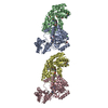
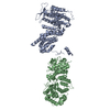
| Movie |
Movie viewer |
|---|---|
| Structure viewer | EM map: SurfViewMolmilJmol/JSmol |
| Supplemental images |

- Downloads & links
-EMDB archive
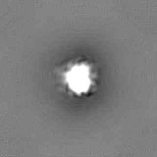
| Map data | emd_25835.map.gz | 115.8 MB | EMDB map data format | |
|---|---|---|---|---|
| Header (meta data) | emd-25835-v30.xmlemd-25835.xml | 20.4 KB 20.4 KB | Display Display | EMDB header |
| FSC (resolution estimation) | emd_25835_fsc.xml | 14.8 KB | Display | FSC data file |
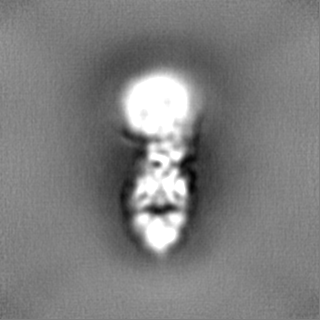
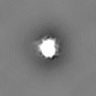
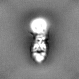
| Images | 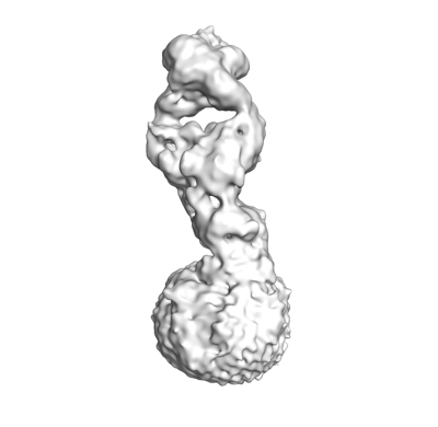 emd_25835.png emd_25835.png | 41.7 KB | ||
| Filedesc metadata | emd-25835.cif.gz | 6.3 KB | ||
| Others | emd_25835_half_map_1.map.gzemd_25835_half_map_2.map.gz | 116 MB 116 MB | ||
| Archive directory |  http://ftp.pdbj.org/pub/emdb/structures/EMD-25835ftp://ftp.pdbj.org/pub/emdb/structures/EMD-25835 http://ftp.pdbj.org/pub/emdb/structures/EMD-25835ftp://ftp.pdbj.org/pub/emdb/structures/EMD-25835 | HTTPS FTP |
-Related structure data
| Related structure data | 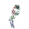 7tdnMC 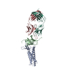 7tdmC M: atomic model generated by this map C: citing same article ( |
|---|---|
| Similar structure data |
-Links
| EMDB pages | EMDB (EBI/PDBe) / EMDataResource |
|---|---|
| Related items in Molecule of the Month |

-Map
| File | Download / File: emd_25835.map.gz / Format: CCP4 / Size: 125 MB / Type: IMAGE STORED AS FLOATING POINT NUMBER (4 BYTES) | ||||||||||||||||||||||||||||||||||||||||||||||||||||||||||||||||||||
|---|---|---|---|---|---|---|---|---|---|---|---|---|---|---|---|---|---|---|---|---|---|---|---|---|---|---|---|---|---|---|---|---|---|---|---|---|---|---|---|---|---|---|---|---|---|---|---|---|---|---|---|---|---|---|---|---|---|---|---|---|---|---|---|---|---|---|---|---|---|
| Voxel size | X=Y=Z: 0.871 Å | ||||||||||||||||||||||||||||||||||||||||||||||||||||||||||||||||||||
| Density |
| ||||||||||||||||||||||||||||||||||||||||||||||||||||||||||||||||||||
| Symmetry | Space group: 1 | ||||||||||||||||||||||||||||||||||||||||||||||||||||||||||||||||||||
| Details | EMDB XML:
CCP4 map header:
| ||||||||||||||||||||||||||||||||||||||||||||||||||||||||||||||||||||
-Supplemental data
-Half map: #2
| File | emd_25835_half_map_1.map | ||||||||||||
|---|---|---|---|---|---|---|---|---|---|---|---|---|---|
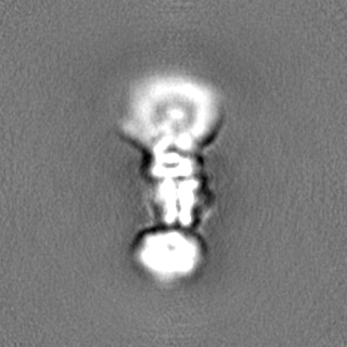
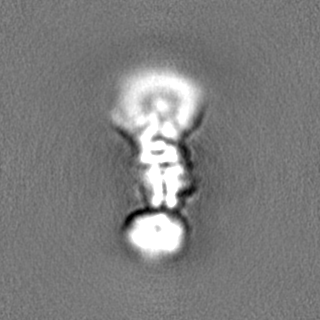
| Projections & Slices |
| ||||||||||||
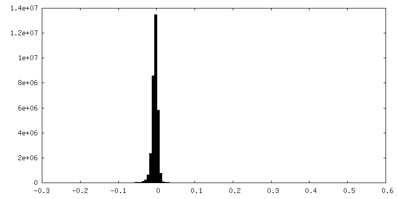
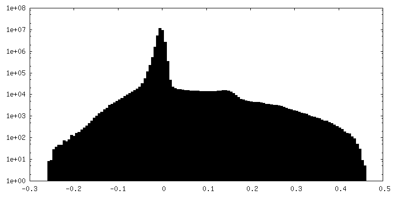
| Density Histograms |
 Z
Z Y
Y X
X


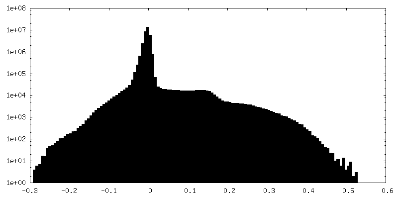
-Half map: #1
| File | emd_25835_half_map_2.map | ||||||||||||
|---|---|---|---|---|---|---|---|---|---|---|---|---|---|
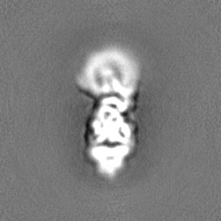
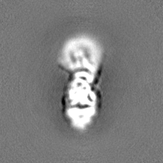
| Projections & Slices |
| ||||||||||||
| Density Histograms |

- Sample components
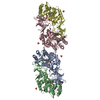
-Entire : Human claudin-4/cCpE/COP-3 Complex
| Entire | Name: Human claudin-4/cCpE/COP-3 Complex |
|---|---|
| Components |
|
-Supramolecule #1: Human claudin-4/cCpE/COP-3 Complex
| Supramolecule | Name: Human claudin-4/cCpE/COP-3 Complex / type: complex / ID: 1 / Parent: 0 / Macromolecule list: all Details: COP-3 sFab fragment bound to Claudin-4/cCpE complex |
|---|---|
| Molecular weight | Theoretical: 85 kDa/nm |
-Macromolecule #1: Claudin-4
| Macromolecule | Name: Claudin-4 / type: protein_or_peptide / ID: 1 / Number of copies: 1 / Enantiomer: LEVO |
|---|---|
| Source (natural) | Organism: Homo sapiens (human) |
| Molecular weight | Theoretical: 22.090201 KDa |
| Recombinant expression | Organism:  Trichoplusia ni (cabbage looper) Trichoplusia ni (cabbage looper) |
| Sequence | String: MASMGLQVMG IALAVLGWLA VMLCCALPMW RVTAFIGSNI VTSQTIWEGL WMNCVVQSTG QMQCKVYDSL LALPQDLQAA RALVIISII VAALGVLLSV VGGKCTNCLE DESAKAKTMI VAGVVFLLAG LMVIVPVSWT AHNIIQDFYN PLVASGQKRE M GASLYVGW ...String: MASMGLQVMG IALAVLGWLA VMLCCALPMW RVTAFIGSNI VTSQTIWEGL WMNCVVQSTG QMQCKVYDSL LALPQDLQAA RALVIISII VAALGVLLSV VGGKCTNCLE DESAKAKTMI VAGVVFLLAG LMVIVPVSWT AHNIIQDFYN PLVASGQKRE M GASLYVGW AASGLLLLGG GLLCCNCPPR TDKPYSAKYS AARSAAASNY V UniProtKB: Claudin-4 |
-Macromolecule #2: Heat-labile enterotoxin B chain
| Macromolecule | Name: Heat-labile enterotoxin B chain / type: protein_or_peptide / ID: 2 / Number of copies: 1 / Enantiomer: LEVO |
|---|---|
| Source (natural) | Organism: Clostridium perfringens (bacteria) |
| Molecular weight | Theoretical: 15.114945 KDa |
| Recombinant expression | Organism: Trichoplusia ni (cabbage looper) |
| Sequence | String: MSTDIEKEIL DLAAATERLN LTDALNSNPA GNLYDWRSSN SYPWTQKLNL HLTITATGQK YRILASKIVD FNIYSNNFNN LVKLEQSLG DGVKDHYVDI SLDAGQYVLV MKANSSYSGN YPYSILFQKF GLVPR UniProtKB: Heat-labile enterotoxin B chain |
-Macromolecule #3: COP-3 Fab Heavy chain
| Macromolecule | Name: COP-3 Fab Heavy chain / type: protein_or_peptide / ID: 3 / Number of copies: 1 / Enantiomer: LEVO |
|---|---|
| Source (natural) | Organism: Homo sapiens (human) |
| Molecular weight | Theoretical: 25.286066 KDa |
| Recombinant expression | Organism: Escherichia coli (E. coli) |
| Sequence | String: EISEVQLVES GGGLVQPGGS LRLSCAASGF NFYSSSIHWV RQAPGKGLEW VAYISSYSGY TYYADSVKGR FTISADTSKN TAYLQMNSL RAEDTAVYYC ARGYGYFDYN FSVGYALDYW GQGTLVTVSS ASTKGPSVFP LAPSSKSTSG GTAALGCLVK D YFPEPVTV ...String: EISEVQLVES GGGLVQPGGS LRLSCAASGF NFYSSSIHWV RQAPGKGLEW VAYISSYSGY TYYADSVKGR FTISADTSKN TAYLQMNSL RAEDTAVYYC ARGYGYFDYN FSVGYALDYW GQGTLVTVSS ASTKGPSVFP LAPSSKSTSG GTAALGCLVK D YFPEPVTV SWNSGALTSG VHTFPAVLQS SGLYSLSSVV TVPSSSLGTQ TYICNVNHKP SNTKVDKKVE PKSCDKTHT |
-Macromolecule #4: COP-3 Fab Light chain
| Macromolecule | Name: COP-3 Fab Light chain / type: protein_or_peptide / ID: 4 / Number of copies: 1 / Enantiomer: LEVO |
|---|---|
| Source (natural) | Organism: Homo sapiens (human) |
| Molecular weight | Theoretical: 23.729328 KDa |
| Recombinant expression | Organism: Escherichia coli (E. coli) |
| Sequence | String: SDIQMTQSPS SLSASVGDRV TITCRASQSV SSAVAWYQQK PGKAPKLLIY SASSLYSGVP SRFSGSRSGT DFTLTISSLQ PEDFATYYC QQSHPWYYPI TFGQGTKVEI KRTVAAPSVF IFPPSDSQLK SGTASVVCLL NNFYPREAKV QWKVDNALQS G NSQESVTE ...String: SDIQMTQSPS SLSASVGDRV TITCRASQSV SSAVAWYQQK PGKAPKLLIY SASSLYSGVP SRFSGSRSGT DFTLTISSLQ PEDFATYYC QQSHPWYYPI TFGQGTKVEI KRTVAAPSVF IFPPSDSQLK SGTASVVCLL NNFYPREAKV QWKVDNALQS G NSQESVTE QDSKDSTYSL SSTLTLSKAD YEKHKVYACE VTHQGLSSPV TKSFNRGEC |
-Experimental details
-Structure determination
| Method | cryo EM |
|---|---|
 Processing Processing | single particle reconstruction |
| Aggregation state | particle |
-Sample preparation
| Concentration | 6.0 mg/mL |
|---|---|
| Buffer | pH: 7.4 |
| Grid | Model: Quantifoil R1.2/1.3 / Mesh: 200 / Pretreatment - Type: GLOW DISCHARGE / Pretreatment - Time: 45 sec. / Pretreatment - Pressure: 0.001 kPa |
| Vitrification | Cryogen name: ETHANE / Chamber humidity: 100 % / Chamber temperature: 277 K / Instrument: FEI VITROBOT MARK IV |
- Electron microscopy
| Microscope | FEI TALOS ARCTICA |
|---|---|
| Electron beam | Acceleration voltage: 200 kV / Electron source: FIELD EMISSION GUN |
| Electron optics | Illumination mode: FLOOD BEAM / Imaging mode: BRIGHT FIELDBright-field microscopy / Nominal defocus max: 2.2 µm / Nominal defocus min: 1.0 µm / Nominal magnification: 120000 |
| Image recording | Film or detector model: FEI FALCON III (4k x 4k) / Detector mode: COUNTING / Number grids imaged: 1 / Number real images: 3758631 / Average electron dose: 39.6 e/Å2 |
| Experimental equipment |  Model: Talos Arctica / Image courtesy: FEI Company |
-Image processing
| Particle selection | Number selected: 305927 |
|---|---|
| Startup model | Type of model: PDB ENTRY PDB model - PDB ID: |
| Initial angle assignment | Type: OTHER |
| Final angle assignment | Type: OTHER |
| Final reconstruction | Number classes used: 1 / Applied symmetry - Point group: C1 (asymmetric) / Resolution.type: BY AUTHOR / Resolution: 5.0 Å / Resolution method: FSC 0.143 CUT-OFF / Software - Name: cryoSPARC / Number images used: 305927 |
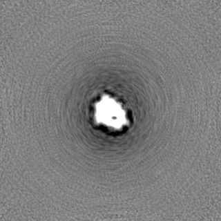
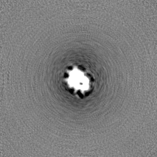
| FSC plot (resolution estimation) | 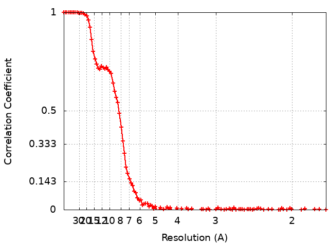 |
-Atomic model buiding 1
| Initial model |
| ||||||||||
|---|---|---|---|---|---|---|---|---|---|---|---|
| Refinement | Space: REAL / Protocol: FLEXIBLE FIT / Overall B value: 413.26 | ||||||||||
| Output model | PDB-7tdn: |
