 Movie
Movie Controller

[English] 日本語
 Yorodumi
- PDB-1sqj: Crystal Structure Analysis of Oligoxyloglucan reducing-end-specif... -
+ Open data

- Basic information
| Entry | Database: PDB / ID: 1sqj | ||||||
|---|---|---|---|---|---|---|---|
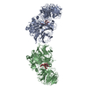
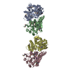
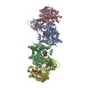
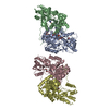
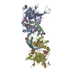
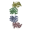
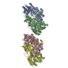
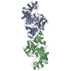
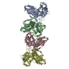
| Title | Crystal Structure Analysis of Oligoxyloglucan reducing-end-specific cellobiohydrolase (OXG-RCBH) | ||||||
 Components Components | oligoxyloglucan reducing-end-specific cellobiohydrolase | ||||||
 Keywords Keywords | HYDROLASE / BETA-PROPELLER | ||||||
| Function / homology |  Function and homology informationoligoxyloglucan reducing-end-specific cellobiohydrolase / oligoxyloglucan reducing-end-specific cellobiohydrolase activity / xyloglucan metabolic process / cellulose catabolic process Function and homology informationoligoxyloglucan reducing-end-specific cellobiohydrolase / oligoxyloglucan reducing-end-specific cellobiohydrolase activity / xyloglucan metabolic process / cellulose catabolic processSimilarity search - Function | ||||||
| Biological species |  Geotrichum sp. M128 (yeast) Geotrichum sp. M128 (yeast) | ||||||
| Method | X-RAY DIFFRACTION / SYNCHROTRON / MAD / Resolution: 2.2 Å | ||||||
 Authors Authors | Yaoi, K. / Kondo, H. / Noro, N. / Suzuki, M. / Tsuda, S. / Mitsuishi, Y. | ||||||
 Citation Citation | Journal: Structure / Year: 2004 Title: Tandem Repeat of a Seven-Bladed beta-Propeller Domain in Oligoxyloglucan Reducing-End-Specific Cellobiohydrolase Authors: Yaoi, K. / Kondo, H. / Noro, N. / Suzuki, M. / Tsuda, S. / Mitsuishi, Y. #1: Journal: J.BIOL.CHEM. / Year: 2002 Title: Purification, characterization, cloning, and expression of a novel xyloglucan-specific glycosidase, oligoxyloglucan reducing end-specific cellobiohydrolase Authors: Yaoi, K. / Mitsuishi, Y. #2: Journal: ACTA CRYSTALLOGR.,SECT.D / Year: 2003 Title: Crystallization and preliminary X-ray crystallographic study on a xyloglucan-specific exo-beta-glycosidase, oligoxyloglucan reducing-end specific cellobiohydrolase Authors: Yaoi, K. / Kondo, H. / Suzuki, M. / Noro, N. / Tsuda, S. / Mitsuishi, Y. | ||||||
| History |
|
- Structure visualization
| Structure viewer | Molecule: MolmilJmol/JSmol |
|---|
- Downloads & links
-Download
| PDBx/mmCIF format | 1sqj.cif.gz | 286.8 KB | Display | PDBx/mmCIF format |
|---|---|---|---|---|
| PDB format | pdb1sqj.ent.gz | 240.8 KB | Display | PDB format |
| PDBx/mmJSON format | 1sqj.json.gz | Tree view | PDBx/mmJSON format | |
| Others |  Other downloads Other downloads |
-Validation report
| Arichive directory | https://data.pdbj.org/pub/pdb/validation_reports/sq/1sqjftp://data.pdbj.org/pub/pdb/validation_reports/sq/1sqj | HTTPS FTP |
|---|
-Related structure data
| Similar structure data |
|---|

-Links
 PDBj
PDBj- Assembly
| Deposited unit | 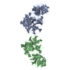
| ||||||||
|---|---|---|---|---|---|---|---|---|---|
| 1 |
| ||||||||
| Unit cell |
|
-Components
| #1: Protein | / OXG-RCBH Mass: 84974.727 Da / Num. of mol.: 2 Source method: isolated from a genetically manipulated source Source: (gene. exp.) Geotrichum sp. M128 (yeast) / Plasmid: pET29a(+) / Production host:  Escherichia coli (E. coli) / Strain (production host): BL21-CodonPlus(DE3)RP Escherichia coli (E. coli) / Strain (production host): BL21-CodonPlus(DE3)RPReferences: UniProt: Q8J0D2, oligoxyloglucan reducing-end-specific cellobiohydrolase#2: Water | ChemComp-HOH / | Water Mass: 18.015 Da / Num. of mol.: 63 / Source method: isolated from a natural source / Formula: H2O Mass: 18.015 Da / Num. of mol.: 63 / Source method: isolated from a natural source / Formula: H2O |
|---|
-Experimental details
-Experiment
| Experiment | Method: X-RAY DIFFRACTION / Number of used crystals: 1 |
|---|
- Sample preparation
| Crystal | Density Matthews: 2.79 Å3/Da / Density % sol: 55.9 % |
|---|---|
| Crystal grow | Temperature: 293 K / Method: vapor diffusion / pH: 5.8 Details: PEG 3000, PEG 400, pH 5.8, VAPOR DIFFUSION, temperature 293K |
-Data collection
| Diffraction | Mean temperature: 95 K | |||||||||||||||
|---|---|---|---|---|---|---|---|---|---|---|---|---|---|---|---|---|
| Diffraction source | Source: SYNCHROTRON / Site: Photon Factory  / Beamline: BL-18B / Wavelength: 0.978, 0.9792, 0.9794, 0.9500 / Beamline: BL-18B / Wavelength: 0.978, 0.9792, 0.9794, 0.9500 | |||||||||||||||
| Detector | Type: ADSC QUANTUM 4 / Detector: CCD / Date: Nov 19, 2002 / Details: mirrors | |||||||||||||||
| Radiation | Monochromator: Si(111) / Protocol: MAD / Monochromatic (M) / Laue (L): M / Scattering type: x-ray | |||||||||||||||
| Radiation wavelength |
| |||||||||||||||
| Reflection | Resolution: 2.2→40 Å / Num. all: 294393 / Num. obs: 294393 / % possible obs: 87.5 % / Observed criterion σ(F): 0 / Observed criterion σ(I): 0 / Redundancy: 3.5 % / Rmerge(I) obs: 0.069 / Net I/σ(I): 9.5 | |||||||||||||||
| Reflection shell | Resolution: 2.2→2.32 Å / Redundancy: 3 % / Rmerge(I) obs: 0.252 / Mean I/σ(I) obs: 3 / % possible all: 70.9 |
- Processing
| Software |
| ||||||||||||||||||||||||||||||||||||||||||||||||||||||||||||||||||||||
|---|---|---|---|---|---|---|---|---|---|---|---|---|---|---|---|---|---|---|---|---|---|---|---|---|---|---|---|---|---|---|---|---|---|---|---|---|---|---|---|---|---|---|---|---|---|---|---|---|---|---|---|---|---|---|---|---|---|---|---|---|---|---|---|---|---|---|---|---|---|---|---|
| Refinement | Method to determine structure: MAD / Resolution: 2.2→20 Å / Cor.coef. Fo:Fc: 0.926 / Cor.coef. Fo:Fc free: 0.901 / SU B: 6.611 / SU ML: 0.166 / Isotropic thermal model: isotropic / Cross valid method: THROUGHOUT / σ(F): 0 / σ(I): 0 / ESU R: 0.301 / ESU R Free: 0.229 / Stereochemistry target values: MAXIMUM LIKELIHOOD
| ||||||||||||||||||||||||||||||||||||||||||||||||||||||||||||||||||||||
| Solvent computation | Ion probe radii: 0.8 Å / Shrinkage radii: 0.8 Å / VDW probe radii: 1.4 Å / Solvent model: BABINET MODEL WITH MASK | ||||||||||||||||||||||||||||||||||||||||||||||||||||||||||||||||||||||
| Displacement parameters | Biso mean: 36.686 Å2
| ||||||||||||||||||||||||||||||||||||||||||||||||||||||||||||||||||||||
| Refinement step | Cycle: LAST / Resolution: 2.2→20 Å
| ||||||||||||||||||||||||||||||||||||||||||||||||||||||||||||||||||||||
| Refine LS restraints |
| ||||||||||||||||||||||||||||||||||||||||||||||||||||||||||||||||||||||
| LS refinement shell | Resolution: 2.2→2.256 Å / Total num. of bins used: 20
|
