 Movie
Movie Controller

[English] 日本語
 Yorodumi
- PDB-7mdh: STRUCTURAL BASIS FOR LIGHT ACITVATION OF A CHLOROPLAST ENZYME. TH... -
+ Open data

- Basic information
| Entry | Database: PDB / ID: 7mdh | ||||||
|---|---|---|---|---|---|---|---|
| Title | STRUCTURAL BASIS FOR LIGHT ACITVATION OF A CHLOROPLAST ENZYME. THE STRUCTURE OF SORGHUM NADP-MALATE DEHYDROGENASE IN ITS OXIDIZED FORM | ||||||
 Components Components | PROTEIN (MALATE DEHYDROGENASE) | ||||||
 Keywords Keywords | CHLOROPLASTIC MALATE DEHYDROGENASE / CHLOROPLASTIC MALATE DEHYDROGENASE (NADP+) / ACTIVATED BY LIGHT | ||||||
| Function / homology |  Function and homology information Function and homology information malate dehydrogenase (NADP+) / malate dehydrogenase (NADP+) activity / malate metabolic process / chloroplast / carbohydrate metabolic process malate dehydrogenase (NADP+) / malate dehydrogenase (NADP+) activity / malate metabolic process / chloroplast / carbohydrate metabolic processSimilarity search - Function | ||||||
| Biological species |  Sorghum bicolor (sorghum) Sorghum bicolor (sorghum) | ||||||
| Method | X-RAY DIFFRACTION / SYNCHROTRON / MOLECULAR REPLACEMENT / Resolution: 2.4 Å | ||||||
 Authors Authors | Johansson, K. / Ramaswamy, S. / Saarinen, M. / Lemaire-Chamley, M. / Issakidis-Bourguet, E. / Miginiac-Maslow, M. / Eklund, H. | ||||||
 Citation Citation | Journal: Biochemistry / Year: 1999 Title: Structural basis for light activation of a chloroplast enzyme: the structure of sorghum NADP-malate dehydrogenase in its oxidized form. Authors: Johansson, K. / Ramaswamy, S. / Saarinen, M. / Lemaire-Chamley, M. / Issakidis-Bourguet, E. / Miginiac-Maslow, M. / Eklund, H. | ||||||
| History |
|
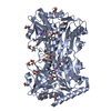
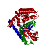
- Structure visualization
| Structure viewer | Molecule: MolmilJmol/JSmol |
|---|
- Downloads & links
-Download
| PDBx/mmCIF format | 7mdh.cif.gz | 283.3 KB | Display | PDBx/mmCIF format |
|---|---|---|---|---|
| PDB format | pdb7mdh.ent.gz | 229.2 KB | Display | PDB format |
| PDBx/mmJSON format | 7mdh.json.gz | Tree view | PDBx/mmJSON format | |
| Others |  Other downloads Other downloads |
-Validation report
| Arichive directory | https://data.pdbj.org/pub/pdb/validation_reports/md/7mdhftp://data.pdbj.org/pub/pdb/validation_reports/md/7mdh | HTTPS FTP |
|---|
-Related structure data
| Related structure data | 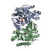 1bmdS S: Starting model for refinement |
|---|---|
| Similar structure data |


-Links
 PDBj
PDBj


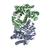
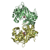
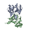
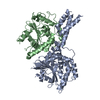
- Assembly
| Deposited unit | 
| ||||||||||||||||
|---|---|---|---|---|---|---|---|---|---|---|---|---|---|---|---|---|---|
| 1 | 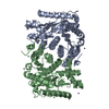
| ||||||||||||||||
| 2 | 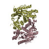
| ||||||||||||||||
| 3 | 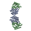
| ||||||||||||||||
| 4 | 
| ||||||||||||||||
| Unit cell |
| ||||||||||||||||
| Noncrystallographic symmetry (NCS) | NCS oper:
|
-Components
| #1: Protein | Mass: 40866.426 Da / Num. of mol.: 4 / Mutation: DELETION OF FIRST 15 N-TERMINAL RESIDUES Source method: isolated from a genetically manipulated source Source: (gene. exp.) Sorghum bicolor (sorghum) / Tissue: LEAF / Organelle: CHLOROPLAST / Plasmid: PET / Species (production host): Escherichia coli / Production host:  Escherichia coli BL21(DE3) (bacteria) / Strain (production host): BL21(DE3) / References: UniProt: P17606, malate dehydrogenase (NADP+) Escherichia coli BL21(DE3) (bacteria) / Strain (production host): BL21(DE3) / References: UniProt: P17606, malate dehydrogenase (NADP+)#2: Chemical | ChemComp-ZN /   Mass: 65.409 Da / Num. of mol.: 17 / Source method: obtained synthetically / Formula: Zn Mass: 65.409 Da / Num. of mol.: 17 / Source method: obtained synthetically / Formula: Zn#3: Water | ChemComp-HOH / | Water Mass: 18.015 Da / Num. of mol.: 265 / Source method: isolated from a natural source / Formula: H2O Mass: 18.015 Da / Num. of mol.: 265 / Source method: isolated from a natural source / Formula: H2O |
|---|
-Experimental details
-Experiment
| Experiment | Method: X-RAY DIFFRACTION / Number of used crystals: 1 |
|---|
- Sample preparation
| Crystal | Density Matthews: 2.74 Å3/Da / Density % sol: 61 % / Description: POLY-ALANINE MODEL | |||||||||||||||
|---|---|---|---|---|---|---|---|---|---|---|---|---|---|---|---|---|
| Crystal grow | pH: 6.5 / Details: pH 6.5 | |||||||||||||||
| Crystal grow | *PLUS Method: unknown | |||||||||||||||
| Components of the solutions | *PLUS
|
-Data collection
| Diffraction | Mean temperature: 100 K |
|---|---|
| Diffraction source | Source: SYNCHROTRON / Site: MAX II  / Beamline: I711 / Wavelength: 0.996 / Beamline: I711 / Wavelength: 0.996 |
| Detector | Type: MAR scanner 345 mm plate / Detector: IMAGE PLATE / Date: Jun 1, 1998 |
| Radiation | Protocol: SINGLE WAVELENGTH / Monochromatic (M) / Laue (L): M / Scattering type: x-ray |
| Radiation wavelength | Wavelength: 0.996 Å / Relative weight: 1 |
| Reflection | Resolution: 2.3→50 Å / Num. obs: 66459 / % possible obs: 94.9 % / Observed criterion σ(I): 0 / Redundancy: 5.2 % / Biso Wilson estimate: 30.2 Å2 / Rsym value: 0.068 / Net I/σ(I): 10.8 |
| Reflection shell | Resolution: 2.4→2.44 Å / % possible all: 99.8 |
| Reflection | *PLUS Rmerge(I) obs: 0.068 |
| Reflection shell | *PLUS % possible obs: 99.8 % / Mean I/σ(I) obs: 4 |
- Processing
| Software |
| ||||||||||||||||||||||||||||||||||||||||||||||||||||||||||||
|---|---|---|---|---|---|---|---|---|---|---|---|---|---|---|---|---|---|---|---|---|---|---|---|---|---|---|---|---|---|---|---|---|---|---|---|---|---|---|---|---|---|---|---|---|---|---|---|---|---|---|---|---|---|---|---|---|---|---|---|---|---|
| Refinement | Method to determine structure: MOLECULAR REPLACEMENT Starting model: 1BMD Resolution: 2.4→50 Å / Rfactor Rfree error: 0.006 / Data cutoff high rms absF: 30748455.68 / Isotropic thermal model: RESTRAINED / Cross valid method: THROUGHOUT / σ(F): 0
| ||||||||||||||||||||||||||||||||||||||||||||||||||||||||||||
| Solvent computation | Solvent model: FLAT MODEL / Bsol: 31 Å2 / ksol: 0.323 e/Å3 | ||||||||||||||||||||||||||||||||||||||||||||||||||||||||||||
| Displacement parameters | Biso mean: 40.3 Å2
| ||||||||||||||||||||||||||||||||||||||||||||||||||||||||||||
| Refine analyze |
| ||||||||||||||||||||||||||||||||||||||||||||||||||||||||||||
| Refinement step | Cycle: LAST / Resolution: 2.4→50 Å
| ||||||||||||||||||||||||||||||||||||||||||||||||||||||||||||
| Refine LS restraints |
| ||||||||||||||||||||||||||||||||||||||||||||||||||||||||||||
| LS refinement shell | Resolution: 2.4→2.44 Å / Rfactor Rfree error: 0.03 / Total num. of bins used: 20
| ||||||||||||||||||||||||||||||||||||||||||||||||||||||||||||
| Xplor file |
| ||||||||||||||||||||||||||||||||||||||||||||||||||||||||||||
| Software | *PLUS Name: CNS / Version: 0.4 / Classification: refinement | ||||||||||||||||||||||||||||||||||||||||||||||||||||||||||||
| Refinement | *PLUS Rfactor obs: 0.221 | ||||||||||||||||||||||||||||||||||||||||||||||||||||||||||||
| Solvent computation | *PLUS | ||||||||||||||||||||||||||||||||||||||||||||||||||||||||||||
| Displacement parameters | *PLUS | ||||||||||||||||||||||||||||||||||||||||||||||||||||||||||||
| Refine LS restraints | *PLUS
| ||||||||||||||||||||||||||||||||||||||||||||||||||||||||||||
| LS refinement shell | *PLUS Rfactor obs: 0.245 |
