 Movie
Movie Controller


+ Open data

- Basic information
| Entry |  | |||||||||
|---|---|---|---|---|---|---|---|---|---|---|
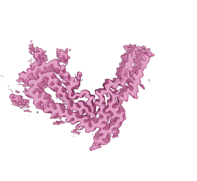
| Title | aRML prion fibril | |||||||||
 Map data Map data | aRML prion EM map | |||||||||
 Sample Sample |
| |||||||||
| Function / homology |  Function and homology information Function and homology informationInsertion of tail-anchored proteins into the endoplasmic reticulum membrane / negative regulation of amyloid precursor protein catabolic process /  lamin binding / regulation of glutamate receptor signaling pathway / regulation of calcium ion import across plasma membrane / aspartic-type endopeptidase inhibitor activity / glycosaminoglycan binding / negative regulation of interleukin-17 production / ATP-dependent protein binding / regulation of potassium ion transmembrane transport ...Insertion of tail-anchored proteins into the endoplasmic reticulum membrane / negative regulation of amyloid precursor protein catabolic process / lamin binding / regulation of glutamate receptor signaling pathway / regulation of calcium ion import across plasma membrane / aspartic-type endopeptidase inhibitor activity / glycosaminoglycan binding / negative regulation of interleukin-17 production / ATP-dependent protein binding / regulation of potassium ion transmembrane transport / negative regulation of dendritic spine maintenance / type 5 metabotropic glutamate receptor binding / cupric ion binding / nucleobase-containing compound metabolic process / response to copper ion / negative regulation of interleukin-2 production / negative regulation of calcineurin-NFAT signaling cascade / negative regulation of T cell receptor signaling pathway / negative regulation of amyloid-beta formation / cuprous ion binding / activation of protein kinase activity / negative regulation of activated T cell proliferation / negative regulation of long-term synaptic potentiation / response to amyloid-beta / negative regulation of type II interferon production / : / intracellular copper ion homeostasis / positive regulation of protein targeting to membrane / response to cadmium ion / side of membrane / inclusion body / regulation of peptidyl-tyrosine phosphorylation / cellular response to copper ion / neuron projection maintenance / molecular condensate scaffold activity / tubulin binding / protein sequestering activity / negative regulation of protein phosphorylation / molecular function activator activity / positive regulation of protein localization to plasma membrane / protein destabilization / protein homooligomerization / negative regulation of DNA-binding transcription factor activity / terminal bouton / cellular response to amyloid-beta / positive regulation of neuron apoptotic process / regulation of protein localization / positive regulation of peptidyl-tyrosine phosphorylation / cellular response to xenobiotic stimulus / signaling receptor activity / amyloid-beta binding / protein-folding chaperone binding / microtubule binding / nuclear membrane / response to oxidative stress / protease binding / mitochondrial outer membrane / transmembrane transporter binding / postsynaptic density / learning or memory / molecular adaptor activity / copper ion binding / membrane raft / intracellular membrane-bounded organelle / dendrite / protein-containing complex binding / negative regulation of apoptotic process / Golgi apparatus / cell surface / endoplasmic reticulum / membrane / identical protein binding / metal ion binding / plasma membrane / cytosol lamin binding / regulation of glutamate receptor signaling pathway / regulation of calcium ion import across plasma membrane / aspartic-type endopeptidase inhibitor activity / glycosaminoglycan binding / negative regulation of interleukin-17 production / ATP-dependent protein binding / regulation of potassium ion transmembrane transport ...Insertion of tail-anchored proteins into the endoplasmic reticulum membrane / negative regulation of amyloid precursor protein catabolic process / lamin binding / regulation of glutamate receptor signaling pathway / regulation of calcium ion import across plasma membrane / aspartic-type endopeptidase inhibitor activity / glycosaminoglycan binding / negative regulation of interleukin-17 production / ATP-dependent protein binding / regulation of potassium ion transmembrane transport / negative regulation of dendritic spine maintenance / type 5 metabotropic glutamate receptor binding / cupric ion binding / nucleobase-containing compound metabolic process / response to copper ion / negative regulation of interleukin-2 production / negative regulation of calcineurin-NFAT signaling cascade / negative regulation of T cell receptor signaling pathway / negative regulation of amyloid-beta formation / cuprous ion binding / activation of protein kinase activity / negative regulation of activated T cell proliferation / negative regulation of long-term synaptic potentiation / response to amyloid-beta / negative regulation of type II interferon production / : / intracellular copper ion homeostasis / positive regulation of protein targeting to membrane / response to cadmium ion / side of membrane / inclusion body / regulation of peptidyl-tyrosine phosphorylation / cellular response to copper ion / neuron projection maintenance / molecular condensate scaffold activity / tubulin binding / protein sequestering activity / negative regulation of protein phosphorylation / molecular function activator activity / positive regulation of protein localization to plasma membrane / protein destabilization / protein homooligomerization / negative regulation of DNA-binding transcription factor activity / terminal bouton / cellular response to amyloid-beta / positive regulation of neuron apoptotic process / regulation of protein localization / positive regulation of peptidyl-tyrosine phosphorylation / cellular response to xenobiotic stimulus / signaling receptor activity / amyloid-beta binding / protein-folding chaperone binding / microtubule binding / nuclear membrane / response to oxidative stress / protease binding / mitochondrial outer membrane / transmembrane transporter binding / postsynaptic density / learning or memory / molecular adaptor activity / copper ion binding / membrane raft / intracellular membrane-bounded organelle / dendrite / protein-containing complex binding / negative regulation of apoptotic process / Golgi apparatus / cell surface / endoplasmic reticulum / membrane / identical protein binding / metal ion binding / plasma membrane / cytosolSimilarity search - Function | |||||||||
| Biological species |  Mus musculus (house mouse) / house mouse (house mouse) Mus musculus (house mouse) / house mouse (house mouse) | |||||||||
| Method | single particle reconstruction / cryo EM / Resolution: 3.0 Å | |||||||||
 Authors Authors | Hoyt F / Standke HG / Artikis E / Caughey B / Kraus A | |||||||||
| Funding support | 2 items
| |||||||||
 Citation Citation | Journal: Mol Cell / Year: 2021 Title: High-resolution structure and strain comparison of infectious mammalian prions. Authors: Allison Kraus / Forrest Hoyt / Cindi L Schwartz / Bryan Hansen / Efrosini Artikis / Andrew G Hughson / Gregory J Raymond / Brent Race / Gerald S Baron / Byron Caughey /  Abstract: Within the extensive range of self-propagating pathologic protein aggregates of mammals, prions are the most clearly infectious (e.g., ∼10 lethal doses per milligram). The structures of such lethal ...Within the extensive range of self-propagating pathologic protein aggregates of mammals, prions are the most clearly infectious (e.g., ∼10 lethal doses per milligram). The structures of such lethal assemblies of PrP molecules have been poorly understood. Here we report a near-atomic core structure of a brain-derived, fully infectious prion (263K strain). Cryo-electron microscopy showed amyloid fibrils assembled with parallel in-register intermolecular β sheets. Each monomer provides one rung of the ordered fibril core, with N-linked glycans and glycolipid anchors projecting outward. Thus, single monomers form the templating surface for incoming monomers at fibril ends, where prion growth occurs. Comparison to another prion strain (aRML) revealed major differences in fibril morphology but, like 263K, an asymmetric fibril cross-section without paired protofilaments. These findings provide structural insights into prion propagation, strains, species barriers, and membrane pathogenesis. This structure also helps frame considerations of factors influencing the relative transmissibility of other pathologic amyloids. | |||||||||
| History |
|
- Structure visualization
| Supplemental images |
|---|
- Downloads & links
-EMDB archive
| Map data | emd_25824.map.gz | 152.7 MB | EMDB map data format | |
|---|---|---|---|---|
| Header (meta data) | emd-25824-v30.xmlemd-25824.xml | 19.4 KB 19.4 KB | Display Display | EMDB header |
| FSC (resolution estimation) | emd_25824_fsc.xml | 13.1 KB | Display | FSC data file |
| Images |  emd_25824.png emd_25824.png | 73.8 KB | ||
| Others | emd_25824_additional_1.map.gzemd_25824_half_map_1.map.gzemd_25824_half_map_2.map.gz | 8.1 MB 153 MB 153 MB | ||
| Archive directory |  http://ftp.pdbj.org/pub/emdb/structures/EMD-25824ftp://ftp.pdbj.org/pub/emdb/structures/EMD-25824 http://ftp.pdbj.org/pub/emdb/structures/EMD-25824ftp://ftp.pdbj.org/pub/emdb/structures/EMD-25824 | HTTPS FTP |
-Related structure data
| Related structure data |  7td6MC M: atomic model generated by this map C: citing same article ( |
|---|---|
| Similar structure data |
-Links
| EMDB pages | EMDB (EBI/PDBe) / EMDataResource |
|---|---|
| Related items in Molecule of the Month |


-Map
| File | Download / File: emd_25824.map.gz / Format: CCP4 / Size: 193.2 MB / Type: IMAGE STORED AS FLOATING POINT NUMBER (4 BYTES) | ||||||||||||||||||||
|---|---|---|---|---|---|---|---|---|---|---|---|---|---|---|---|---|---|---|---|---|---|
| Annotation | aRML prion EM map | ||||||||||||||||||||
| Voxel size | X=Y=Z: 1.09 Å | ||||||||||||||||||||
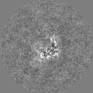
| Density |
| ||||||||||||||||||||
| Symmetry | Space group: 1 | ||||||||||||||||||||
| Details | EMDB XML:
|
-Supplemental data
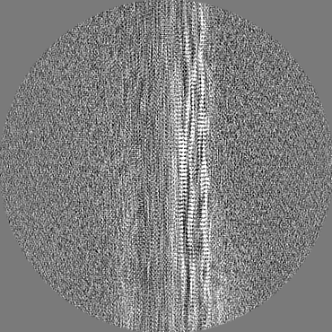
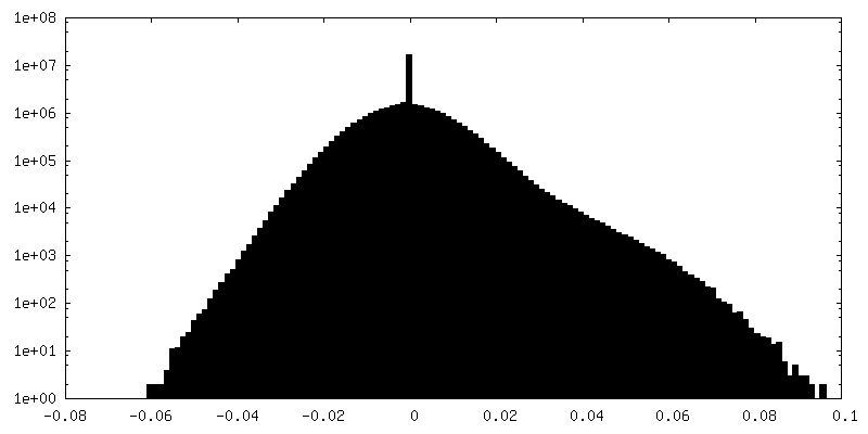
-Additional map: aRML prion EM post-processed masked map out of Relion
| File | emd_25824_additional_1.map | ||||||||||||
|---|---|---|---|---|---|---|---|---|---|---|---|---|---|
| Annotation | aRML prion EM post-processed masked map out of Relion | ||||||||||||
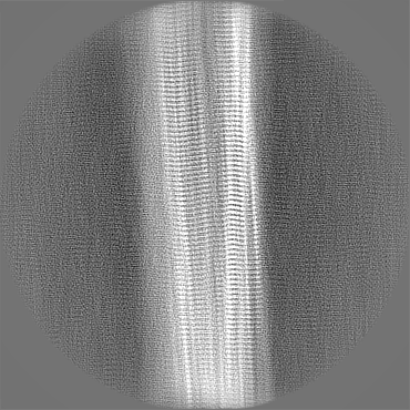
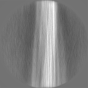
| Projections & Slices |
| ||||||||||||
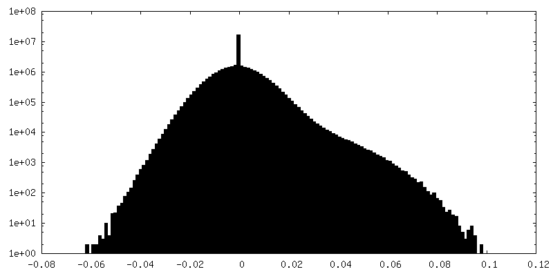
| Density Histograms |
 Z
Z Y
Y X
X


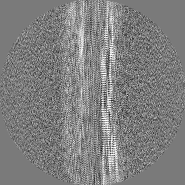
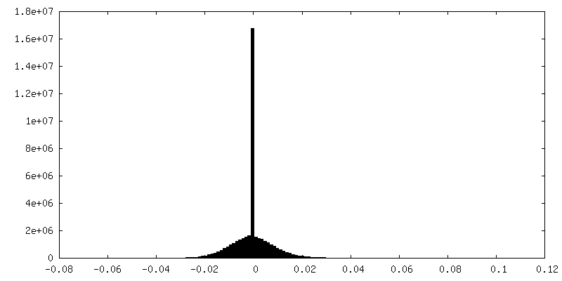
-Half map: aRML prion refinement half-map 1
| File | emd_25824_half_map_1.map | ||||||||||||
|---|---|---|---|---|---|---|---|---|---|---|---|---|---|
| Annotation | aRML prion refinement half-map 1 | ||||||||||||
| Projections & Slices |
| ||||||||||||
| Density Histograms |


-Half map: aRML prion refinement half-map 2
| File | emd_25824_half_map_2.map | ||||||||||||
|---|---|---|---|---|---|---|---|---|---|---|---|---|---|
| Annotation | aRML prion refinement half-map 2 | ||||||||||||
| Projections & Slices |
| ||||||||||||
| Density Histograms |


- Sample components
-Entire : aRML prion
| Entire | Name: aRML prion |
|---|---|
| Components |
|
-Supramolecule #1: aRML prion
| Supramolecule | Name: aRML prion / type: complex / Chimera: Yes / ID: 1 / Parent: 0 / Macromolecule list: all / Details: anchorless RML prion fibril |
|---|---|
| Source (natural) | Organism: Mus musculus (house mouse) / Tissue: brain-derived |
-Macromolecule #1: Major prion protein
| Macromolecule | Name: Major prion protein / type: protein_or_peptide / ID: 1 Details: proteinase-K resistant aRML prion fibril ordered core encompasses residues 93-230 Number of copies: 5 / Enantiomer: LEVO |
|---|---|
| Source (natural) | Organism: house mouse (house mouse) |
| Molecular weight | Theoretical: 28.008393 KDa |
| Sequence | String: MANLGYWLLA LFVTMWTDVG LCKKRPKPGG WNTGGSRYPG QGSPGGNRYP PQGGTWGQPH GGGWGQPHGG SWGQPHGGSW GQPHGGGWG QGGGTHNQWN KPSKPKTNLK HVAGAAAAGA VVGGLGGYML GSAMSRPMIH FGNDWEDRYY RENMYRYPNQ V YYRPVDQY ...String: MANLGYWLLA LFVTMWTDVG LCKKRPKPGG WNTGGSRYPG QGSPGGNRYP PQGGTWGQPH GGGWGQPHGG SWGQPHGGSW GQPHGGGWG QGGGTHNQWN KPSKPKTNLK HVAGAAAAGA VVGGLGGYML GSAMSRPMIH FGNDWEDRYY RENMYRYPNQ V YYRPVDQY SNQNNFVHDC VNITIKQHTV TTTTKGENFT ETDVKMMERV VEQMCVTQYQ KESQAYYDGR RSSSTVLFSS PP VILLISF LIFLIVG |
-Experimental details
-Structure determination
| Method | cryo EM |
|---|---|
 Processing Processing | single particle reconstruction |
| Aggregation state | particle |
-Sample preparation
| Concentration | 0.4 mg/mL |
|---|---|
| Buffer | pH: 7.4 |
| Grid | Model: C-flat-1.2/1.3 / Material: COPPER / Mesh: 300 / Pretreatment - Type: GLOW DISCHARGE |
| Vitrification | Cryogen name: ETHANE |
| Details | purified aRML prion fibrils |
- Electron microscopy
| Microscope | FEI TITAN KRIOS |
|---|---|
| Electron beam | Acceleration voltage: 300 kV / Electron source: FIELD EMISSION GUN |
| Electron optics | Illumination mode: FLOOD BEAM / Imaging mode: BRIGHT FIELDBright-field microscopy / Nominal defocus max: 2.7 µm / Nominal defocus min: 0.5 µm / Nominal magnification: 81000 |
| Sample stage | Specimen holder model: FEI TITAN KRIOS AUTOGRID HOLDER / Cooling holder cryogen: NITROGEN |
| Image recording | Film or detector model: GATAN K3 BIOQUANTUM (6k x 4k) / Average electron dose: 60.0 e/Å2 |
| Experimental equipment |  Model: Titan Krios / Image courtesy: FEI Company |
-Image processing
| Particle selection | Number selected: 135939 |
|---|---|
| CTF correction | Software - Name: CTFFIND (ver. 4.1) |
| Initial angle assignment | Type: MAXIMUM LIKELIHOOD / Software - Name: RELION (ver. 3.1) |
| Final angle assignment | Type: MAXIMUM LIKELIHOOD / Software - Name: RELION (ver. 3.1) |
| Final reconstruction | Applied symmetry - Point group: C1 (asymmetric) / Resolution.type: BY AUTHOR / Resolution: 3.0 Å / Resolution method: FSC 0.143 CUT-OFF / Number images used: 15857 |
| FSC plot (resolution estimation) |  |
