 Movie
Movie Controller

[English] 日本語
 Yorodumi
- EMDB-24802: Wild-type Escherichia coli stalled ribosome with antibiotic radezolid -
+ Open data

- Basic information
| Entry | Database: EMDB / ID: EMD-24802 | ||||||||||||||||||||||||||||||||||||
|---|---|---|---|---|---|---|---|---|---|---|---|---|---|---|---|---|---|---|---|---|---|---|---|---|---|---|---|---|---|---|---|---|---|---|---|---|---|
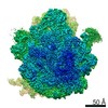
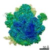
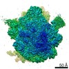
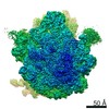
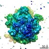
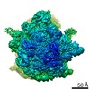
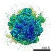
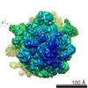
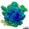
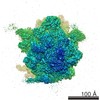
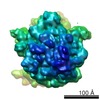
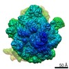
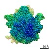
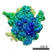
| Title | Wild-type Escherichia coli stalled ribosome with antibiotic radezolid | ||||||||||||||||||||||||||||||||||||
 Map data Map data | final unsharpened map used for model building | ||||||||||||||||||||||||||||||||||||
 Sample Sample |
| ||||||||||||||||||||||||||||||||||||
 Keywords Keywords |  stalled ribosome / oxazolidinone / radezolid / RIBOSOME stalled ribosome / oxazolidinone / radezolid / RIBOSOME | ||||||||||||||||||||||||||||||||||||
| Function / homology |  Function and homology information Function and homology informationmisfolded RNA binding / Group I intron splicing / RNA folding / positive regulation of ribosome biogenesis / DnaA-L2 complex / negative regulation of translational initiation / negative regulation of DNA-templated DNA replication initiation / mRNA regulatory element binding translation repressor activity / assembly of large subunit precursor of preribosome / positive regulation of RNA splicing ...misfolded RNA binding / Group I intron splicing / RNA folding / positive regulation of ribosome biogenesis / DnaA-L2 complex / negative regulation of translational initiation / negative regulation of DNA-templated DNA replication initiation / mRNA regulatory element binding translation repressor activity / assembly of large subunit precursor of preribosome / positive regulation of RNA splicing / cytosolic ribosome assembly / transcription antitermination / regulation of cell growth / DNA-templated transcription termination / maintenance of translational fidelity / ribosomal large subunit assembly / mRNA 5'-UTR binding / ribosomal small subunit biogenesis / ribosomal small subunit assembly / small ribosomal subunit rRNA binding / large ribosomal subunit / ribosome binding / 5S rRNA binding / large ribosomal subunit rRNA binding / small ribosomal subunit / cytosolic small ribosomal subunit / transferase activity / cytosolic large ribosomal subunit / cytoplasmic translation / tRNA binding / negative regulation of translation / rRNA binding / ribosome / structural constituent of ribosome / ribonucleoprotein complex / translation / response to antibiotic / mRNA binding / RNA binding / zinc ion binding / membrane / metal ion binding / cytosol / cytoplasmSimilarity search - Function | ||||||||||||||||||||||||||||||||||||
| Biological species |  Escherichia coli (E. coli) Escherichia coli (E. coli) | ||||||||||||||||||||||||||||||||||||
| Method | single particle reconstruction / cryo EM / Resolution: 2.48 Å | ||||||||||||||||||||||||||||||||||||
 Authors Authors | Young ID / Stojkovic V / Tsai K / Lee DJ | ||||||||||||||||||||||||||||||||||||
| Funding support | 11 items
| ||||||||||||||||||||||||||||||||||||
 Citation Citation | Journal: Nat Struct Mol Biol / Year: 2022 Title: Structural basis for context-specific inhibition of translation by oxazolidinone antibiotics. Authors: Kaitlyn Tsai / Vanja Stojković / D John Lee / Iris D Young / Teresa Szal / Dorota Klepacki / Nora Vázquez-Laslop / Alexander S Mankin / James S Fraser / Danica Galonić Fujimori /  Abstract: The antibiotic linezolid, the first clinically approved member of the oxazolidinone class, inhibits translation of bacterial ribosomes by binding to the peptidyl transferase center. Recent work has ...The antibiotic linezolid, the first clinically approved member of the oxazolidinone class, inhibits translation of bacterial ribosomes by binding to the peptidyl transferase center. Recent work has demonstrated that linezolid does not inhibit peptide bond formation at all sequences but rather acts in a context-specific manner, namely when alanine occupies the penultimate position of the nascent chain. However, the molecular basis for context-specificity has not been elucidated. Here we show that the second-generation oxazolidinone radezolid also induces stalling with a penultimate alanine, and we determine high-resolution cryo-EM structures of linezolid- and radezolid-stalled ribosome complexes to explain their mechanism of action. These structures reveal that the alanine side chain fits within a small hydrophobic crevice created by oxazolidinone, resulting in improved ribosome binding. Modification of the ribosome by the antibiotic resistance enzyme Cfr disrupts stalling due to repositioning of the modified nucleotide. Together, our findings provide molecular understanding for the context-specificity of oxazolidinones. | ||||||||||||||||||||||||||||||||||||
| History |
|
- Structure visualization
| Movie |
Movie viewer |
|---|---|
| Structure viewer | EM map: SurfViewMolmilJmol/JSmol |
| Supplemental images |

- Downloads & links
-EMDB archive
| Map data | emd_24802.map.gz | 474.1 MB | EMDB map data format | |
|---|---|---|---|---|
| Header (meta data) | emd-24802-v30.xmlemd-24802.xml | 70.8 KB 70.8 KB | Display Display | EMDB header |
| FSC (resolution estimation) | emd_24802_fsc.xml | 23.2 KB | Display | FSC data file |
| Images | 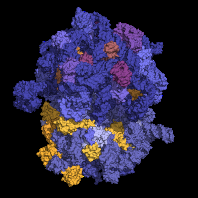 emd_24802.png emd_24802.png | 155.7 KB | ||
| Filedesc metadata | emd-24802.cif.gz | 14.5 KB | ||
| Archive directory |  http://ftp.pdbj.org/pub/emdb/structures/EMD-24802ftp://ftp.pdbj.org/pub/emdb/structures/EMD-24802 http://ftp.pdbj.org/pub/emdb/structures/EMD-24802ftp://ftp.pdbj.org/pub/emdb/structures/EMD-24802 | HTTPS FTP |
-Related structure data
| Related structure data |  7s1iMC  7s1gC  7s1hC  7s1jC  7s1kC M: atomic model generated by this map C: citing same article ( |
|---|---|
| Similar structure data |
-Links
| EMDB pages | EMDB (EBI/PDBe) / EMDataResource |
|---|---|
| Related items in Molecule of the Month |


-Map
| File | Download / File: emd_24802.map.gz / Format: CCP4 / Size: 512 MB / Type: IMAGE STORED AS FLOATING POINT NUMBER (4 BYTES) | ||||||||||||||||||||||||||||||||||||||||||||||||||||||||||||||||||||
|---|---|---|---|---|---|---|---|---|---|---|---|---|---|---|---|---|---|---|---|---|---|---|---|---|---|---|---|---|---|---|---|---|---|---|---|---|---|---|---|---|---|---|---|---|---|---|---|---|---|---|---|---|---|---|---|---|---|---|---|---|---|---|---|---|---|---|---|---|---|
| Annotation | final unsharpened map used for model building | ||||||||||||||||||||||||||||||||||||||||||||||||||||||||||||||||||||
| Voxel size | X=Y=Z: 0.825 Å | ||||||||||||||||||||||||||||||||||||||||||||||||||||||||||||||||||||
| Density |
| ||||||||||||||||||||||||||||||||||||||||||||||||||||||||||||||||||||
| Symmetry | Space group: 1 | ||||||||||||||||||||||||||||||||||||||||||||||||||||||||||||||||||||
| Details | EMDB XML:
CCP4 map header:
| ||||||||||||||||||||||||||||||||||||||||||||||||||||||||||||||||||||
-Supplemental data
- Sample components
+Entire : wild-type Escherichia coli stalled ribosome with antibiotic radezolid
+Supramolecule #1: wild-type Escherichia coli stalled ribosome with antibiotic radezolid
+Macromolecule #1: 30S ribosomal protein S19
+Macromolecule #2: 30S ribosomal protein S20
+Macromolecule #3: 30S ribosomal protein S21
+Macromolecule #7: 30S ribosomal protein S2
+Macromolecule #8: 30S ribosomal protein S3
+Macromolecule #9: 30S ribosomal protein S4
+Macromolecule #10: 30S ribosomal protein S5
+Macromolecule #11: 30S ribosomal protein S6
+Macromolecule #14: 50S ribosomal protein L2
+Macromolecule #15: 50S ribosomal protein L3
+Macromolecule #16: 50S ribosomal protein L4
+Macromolecule #17: 50S ribosomal protein L5
+Macromolecule #18: 50S ribosomal protein L6
+Macromolecule #19: 50S ribosomal protein L9
+Macromolecule #20: 50S ribosomal protein L31
+Macromolecule #21: 50S ribosomal protein L13
+Macromolecule #22: 50S ribosomal protein L14
+Macromolecule #23: 50S ribosomal protein L15
+Macromolecule #24: 50S ribosomal protein L16
+Macromolecule #25: 50S ribosomal protein L17
+Macromolecule #26: 50S ribosomal protein L18
+Macromolecule #27: 50S ribosomal protein L19
+Macromolecule #28: 50S ribosomal protein L20
+Macromolecule #29: Ribosomal protein L21
+Macromolecule #30: 50S ribosomal protein L22
+Macromolecule #31: 50S ribosomal protein L23
+Macromolecule #32: 50S ribosomal protein L24
+Macromolecule #33: 50S ribosomal protein L25
+Macromolecule #34: 50S ribosomal protein L27
+Macromolecule #35: 50S ribosomal protein L28
+Macromolecule #36: 50S ribosomal protein L29
+Macromolecule #37: 50S ribosomal protein L30
+Macromolecule #38: 50S ribosomal protein L32
+Macromolecule #39: 50S ribosomal protein L33
+Macromolecule #40: 50S ribosomal protein L34
+Macromolecule #41: 50S ribosomal protein L35
+Macromolecule #42: 50S ribosomal protein L36
+Macromolecule #43: 30S ribosomal protein S7
+Macromolecule #44: 30S ribosomal protein S8
+Macromolecule #45: 30S ribosomal protein S9
+Macromolecule #46: 30S ribosomal protein S10
+Macromolecule #47: 30S ribosomal protein S11
+Macromolecule #48: nascent peptide chain
+Macromolecule #49: 30S ribosomal protein S12
+Macromolecule #50: 30S ribosomal protein S13
+Macromolecule #51: 30S ribosomal protein S14
+Macromolecule #52: 30S ribosomal protein S15
+Macromolecule #53: 30S ribosomal protein S16
+Macromolecule #54: 30S ribosomal protein S17
+Macromolecule #55: 30S ribosomal protein S18
+Macromolecule #4: mRNA
+Macromolecule #5: tRNA(PHE)
+Macromolecule #6: 16S rRNA
+Macromolecule #12: 23S rRNA
+Macromolecule #13: 5S rRNA
+Macromolecule #56: MAGNESIUM ION
+Macromolecule #57: Radezolid

+Macromolecule #58: ZINC ION
+Macromolecule #59: water

-Experimental details
-Structure determination
| Method | cryo EM |
|---|---|
 Processing Processing | single particle reconstruction |
| Aggregation state | particle |
-Sample preparation
| Buffer | pH: 7.5 |
|---|---|
| Grid | Model: Quantifoil R1.2/1.3 / Material: COPPER / Mesh: 300 / Support film - Material: CARBON / Support film - topology: CONTINUOUS / Support film - Film thickness: 2 / Pretreatment - Type: GLOW DISCHARGE / Pretreatment - Time: 30 sec. / Details: 15 mA |
| Vitrification | Cryogen name: ETHANE / Chamber humidity: 95 % / Chamber temperature: 283.2 K / Instrument: FEI VITROBOT MARK IV |
- Electron microscopy
| Microscope | FEI TITAN KRIOS |
|---|---|
| Electron beam | Acceleration voltage: 300 kV / Electron source: FIELD EMISSION GUN |
| Electron optics | Illumination mode: FLOOD BEAM / Imaging mode: BRIGHT FIELDBright-field microscopy / Cs: 2.7 mm / Nominal defocus max: 1.2 µm / Nominal defocus min: 0.5 µm |
| Specialist optics | Energy filter - Slit width: 20 eV |
| Sample stage | Specimen holder model: FEI TITAN KRIOS AUTOGRID HOLDER / Cooling holder cryogen: NITROGEN |
| Image recording | Film or detector model: GATAN K2 SUMMIT (4k x 4k) / Detector mode: SUPER-RESOLUTION / Number grids imaged: 1 / Number real images: 3566 / Average electron dose: 52.8 e/Å2 |
| Experimental equipment |  Model: Titan Krios / Image courtesy: FEI Company |
-Image processing
| Particle selection | Number selected: 292882 |
|---|---|
| Startup model | Type of model: NONE |
| Initial angle assignment | Type: PROJECTION MATCHING / Software - Name: cisTEM (ver. v1.0.0-beta) |
| Final 3D classification | Number classes: 1 / Software - Name: cisTEM (ver. v1.0.0-beta) |
| Final angle assignment | Type: PROJECTION MATCHING / Software - Name: cisTEM (ver. v1.0.0-beta) |
| Final reconstruction | Applied symmetry - Point group: C1 (asymmetric) / Algorithm: BACK PROJECTION / Resolution.type: BY AUTHOR / Resolution: 2.48 Å / Resolution method: FSC 0.143 CUT-OFF / Software - Name: cisTEM (ver. v1.0.0-beta) / Number images used: 240596 |
| FSC plot (resolution estimation) | 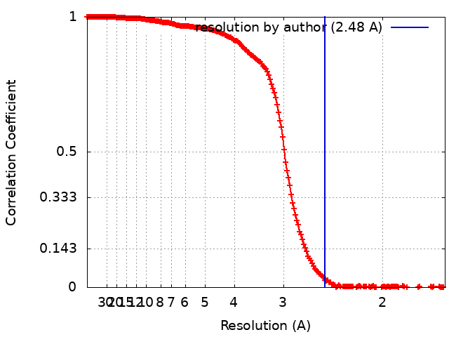 |
-Atomic model buiding 1
| Refinement | Space: REAL / Protocol: FLEXIBLE FIT |
|---|---|
| Output model | PDB-7s1i: |
