National Institutes of Health/National Cancer Institute (NIH/NCI)
R35 CA197582
United States
National Institutes of Health/National Cancer Institute (NIH/NCI)
R50 CA243899
United States
National Natural Science Foundation of China (NSFC)
81872915
China
National Natural Science Foundation of China (NSFC)
82073904
China
National Natural Science Foundation of China (NSFC)
81922071
China
National Natural Science Foundation of China (NSFC)
81773792
China
National Natural Science Foundation of China (NSFC)
81973373
China
National Natural Science Foundation of China (NSFC)
21704064
China
Ministry of Science and Technology (MoST, China)
2018ZX09735 001
China
Ministry of Science and Technology (MoST, China)
2018ZX09711002 002 005
China
Ministry of Science and Technology (MoST, China)
2018YFA0507000
China
Ministry of Science and Technology (MoST, China)
2019YFA0508800
China
Chinese Academy of Sciences
NNCAS 2017 1 CC
China
Citation
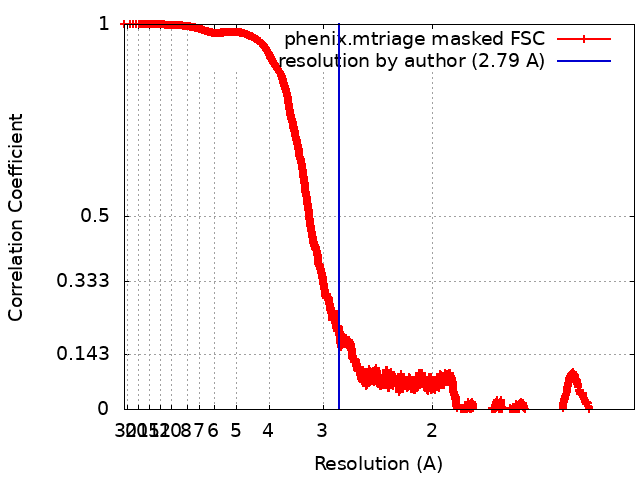
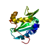
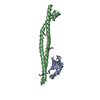
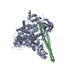
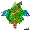
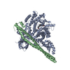
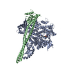
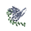
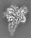
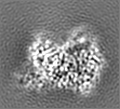
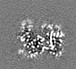
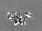
Journal: Proc Natl Acad Sci U S A / Year: 2021 Title: Cryo-EM structures of PI3Kα reveal conformational changes during inhibition and activation. Authors: Xiao Liu / Su Yang / Jonathan R Hart / Yingna Xu / Xinyu Zou / Huibing Zhang / Qingtong Zhou / Tian Xia / Yan Zhang / Dehua Yang / Ming-Wei Wang / Peter K Vogt / Abstract: Phosphoinositide 3-kinases (PI3Ks) are lipid kinases essential for growth and metabolism. Their aberrant activation is associated with many types of cancers. Here we used single-particle cryoelectron ...Phosphoinositide 3-kinases (PI3Ks) are lipid kinases essential for growth and metabolism. Their aberrant activation is associated with many types of cancers. Here we used single-particle cryoelectron microscopy (cryo-EM) to determine three distinct conformations of full-length PI3Kα (p110α-p85α): the unliganded heterodimer PI3Kα, PI3Kα bound to the p110α-specific inhibitor BYL-719, and PI3Kα exposed to an activating phosphopeptide. The cryo-EM structures of unbound and of BYL-719-bound PI3Kα are in general accord with published crystal structures. Local deviations are presented and discussed. BYL-719 stabilizes the structure of PI3Kα, but three regions of low-resolution extra density remain and are provisionally assigned to the cSH2, BH, and SH3 domains of p85. One of the extra density regions is in contact with the kinase domain blocking access to the catalytic site. This conformational change indicates that the effects of BYL-719 on PI3Kα activity extend beyond competition with adenosine triphosphate (ATP). In unliganded PI3Kα, the DFG motif occurs in the "in" and "out" positions. In BYL-719-bound PI3Kα, only the DFG-in position, corresponding to the active conformation of the kinase, was observed. The phosphopeptide-bound structure of PI3Kα is composed of a stable core resolved at 3.8 Å. It contains all p110α domains except the adaptor-binding domain (ABD). The p85α domains, linked to the core through the ABD, are no longer resolved, implying that the phosphopeptide activates PI3Kα by fully releasing the niSH2 domain from binding to p110α. The structures presented here show the basal form of the full-length PI3Kα dimer and document conformational changes related to the activated and inhibited states.
In the structure databanks used in Yorodumi, some data are registered as the other names, "COVID-19 virus" and "2019-nCoV". Here are the details of the virus and the list of structure data.
Jan 31, 2019. EMDB accession codes are about to change! (news from PDBe EMDB page)
EMDB accession codes are about to change! (news from PDBe EMDB page)
The allocation of 4 digits for EMDB accession codes will soon come to an end. Whilst these codes will remain in use, new EMDB accession codes will include an additional digit and will expand incrementally as the available range of codes is exhausted. The current 4-digit format prefixed with “EMD-” (i.e. EMD-XXXX) will advance to a 5-digit format (i.e. EMD-XXXXX), and so on. It is currently estimated that the 4-digit codes will be depleted around Spring 2019, at which point the 5-digit format will come into force.
The EM Navigator/Yorodumi systems omit the EMD- prefix.
Related info.:Q: What is EMD? / ID/Accession-code notation in Yorodumi/EM Navigator
Yorodumi is a browser for structure data from EMDB, PDB, SASBDB, etc.
This page is also the successor to EM Navigator detail page, and also detail information page/front-end page for Omokage search.
The word "yorodu" (or yorozu) is an old Japanese word meaning "ten thousand". "mi" (miru) is to see.
Related info.:EMDB / PDB / SASBDB / Comparison of 3 databanks / Yorodumi Search / Aug 31, 2016. New EM Navigator & Yorodumi / Yorodumi Papers / Jmol/JSmol / Function and homology information / Changes in new EM Navigator and Yorodumi
 Movie
Movie Controller


 Open data
 Basic information
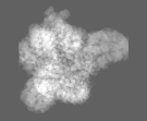
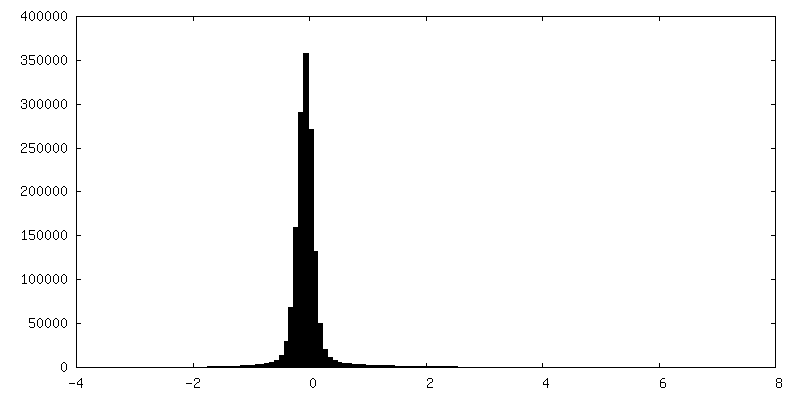
 Map data
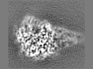
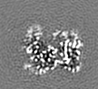
 Sample
 Function and homology information
 transmembrane receptor protein tyrosine kinase adaptor activity / RHOD GTPase cycle / positive regulation of endoplasmic reticulum unfolded protein response / Signaling by cytosolic FGFR1 fusion mutants /
 Authors
 United States,
  China, 13 items
  Citation
 Structure visualization
 Downloads & links
 emd_24081.png
 http://ftp.pdbj.org/pub/emdb/structures/EMD-24081


 Z
Z Y
Y X
X


 Sample components

 Processing
 Electron microscopy