 Movie
Movie Controller

[English] 日本語
 Yorodumi
- EMDB-18651: GDNF/GFRa1 cell adhesion complex bridging between adhering liposomes. -
+ Open data

- Basic information
| Entry |  | |||||||||
|---|---|---|---|---|---|---|---|---|---|---|
| Title | GDNF/GFRa1 cell adhesion complex bridging between adhering liposomes. | |||||||||
 Map data Map data | Reconstructed tomogram of zGDNF-zGFRa1 complexes bridging between adhering liposomes. Tomogram reconstructions were generated with a SIRT-like filter with 5 iterations and binned by 4. | |||||||||
 Sample Sample |
| |||||||||
 Keywords Keywords | Synaptic /  Adhesion / Complex / LiCAM / CELL ADHESION Adhesion / Complex / LiCAM / CELL ADHESION | |||||||||
| Function / homology |  Function and homology information Function and homology informationRAF/MAP kinase cascade / : / diencephalon development / positive regulation of ureteric bud formation / postganglionic parasympathetic fiber development / positive regulation of monooxygenase activity / regulation of dopaminergic neuron differentiation / glial cell-derived neurotrophic factor receptor binding / regulation of morphogenesis of a branching structure / regulation of dopamine uptake involved in synaptic transmission ...RAF/MAP kinase cascade / : / diencephalon development / positive regulation of ureteric bud formation / postganglionic parasympathetic fiber development / positive regulation of monooxygenase activity / regulation of dopaminergic neuron differentiation / glial cell-derived neurotrophic factor receptor binding / regulation of morphogenesis of a branching structure / regulation of dopamine uptake involved in synaptic transmission / enteric nervous system development / positive regulation of branching involved in ureteric bud morphogenesis / peristalsis / sympathetic nervous system development / peripheral nervous system development / mRNA stabilization / metanephros development / neural crest cell migration / branching involved in ureteric bud morphogenesis / MAP kinase kinase kinase activity / growth factor activity / receptor tyrosine kinase binding / neuron projection development / signaling receptor activity / nervous system development / protein-containing complex assembly / negative regulation of neuron apoptotic process / receptor complex / external side of plasma membrane / protein homodimerization activity / positive regulation of transcription by RNA polymerase II / extracellular space / extracellular regionSimilarity search - Function | |||||||||
| Biological species |  Danio rerio (zebrafish) Danio rerio (zebrafish) | |||||||||
| Method | electron tomography / cryo EM | |||||||||
 Authors Authors | Houghton FM / Briggs DC / McDonald NQ | |||||||||
| Funding support |  United Kingdom, 1 items United Kingdom, 1 items
| |||||||||
 Citation Citation | Journal: Nat Commun / Year: 2023 Title: Architecture and regulation of a GDNF-GFRα1 synaptic adhesion assembly. Authors: F M Houghton / S E Adams / A S Ríos / L Masino / A G Purkiss / D C Briggs / F Ledda / N Q McDonald /  Abstract: Glial-cell line derived neurotrophic factor (GDNF) bound to its co-receptor GFRα1 stimulates the RET receptor tyrosine kinase, promoting neuronal survival and neuroprotection. The GDNF-GFRα1 ...Glial-cell line derived neurotrophic factor (GDNF) bound to its co-receptor GFRα1 stimulates the RET receptor tyrosine kinase, promoting neuronal survival and neuroprotection. The GDNF-GFRα1 complex also supports synaptic cell adhesion independently of RET. Here, we describe the structure of a decameric GDNF-GFRα1 assembly determined by crystallography and electron microscopy, revealing two GFRα1 pentamers bridged by five GDNF dimers. We reconsitituted the assembly between adhering liposomes and used cryo-electron tomography to visualize how the complex fulfils its membrane adhesion function. The GFRα1:GFRα1 pentameric interface was further validated both in vitro by native PAGE and in cellulo by cell-clustering and dendritic spine assays. Finally, we provide biochemical and cell-based evidence that RET and heparan sulfate cooperate to prevent assembly of the adhesion complex by competing for the adhesion interface. Our results provide a mechanistic framework to understand GDNF-driven cell adhesion, its relationship to trophic signalling, and the central role played by GFRα1. | |||||||||
| History |
|
- Structure visualization
| Supplemental images |
|---|

- Downloads & links
-EMDB archive
| Map data | emd_18651.map.gz | 10.3 MB | EMDB map data format | |
|---|---|---|---|---|
| Header (meta data) | emd-18651-v30.xmlemd-18651.xml | 13.1 KB 13.1 KB | Display Display | EMDB header |
| Images | 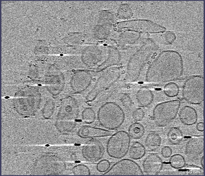 emd_18651.png emd_18651.png | 181.6 KB | ||
| Filedesc metadata | emd-18651.cif.gz | 5.1 KB | ||
| Archive directory |  http://ftp.pdbj.org/pub/emdb/structures/EMD-18651ftp://ftp.pdbj.org/pub/emdb/structures/EMD-18651 http://ftp.pdbj.org/pub/emdb/structures/EMD-18651ftp://ftp.pdbj.org/pub/emdb/structures/EMD-18651 | HTTPS FTP |
-Related structure data
-Links
| EMDB pages | EMDB (EBI/PDBe) / EMDataResource |
|---|---|
| Related items in Molecule of the Month |


-Map
| File | Download / File: emd_18651.map.gz / Format: CCP4 / Size: 21.8 MB / Type: IMAGE STORED AS SIGNED BYTE | ||||||||||||||||||||
|---|---|---|---|---|---|---|---|---|---|---|---|---|---|---|---|---|---|---|---|---|---|
| Annotation | Reconstructed tomogram of zGDNF-zGFRa1 complexes bridging between adhering liposomes. Tomogram reconstructions were generated with a SIRT-like filter with 5 iterations and binned by 4. | ||||||||||||||||||||
| Voxel size | X=Y=Z: 13.02 Å | ||||||||||||||||||||
| Density |
| ||||||||||||||||||||
| Symmetry | Space group: 1 | ||||||||||||||||||||
| Details | EMDB XML:
|
-Supplemental data
- Sample components
-Entire : GDNF/GFRa1 cell adhesion complex assembled onto liposomes.
| Entire | Name: GDNF/GFRa1 cell adhesion complex assembled onto liposomes. |
|---|---|
| Components |
|
-Supramolecule #1: GDNF/GFRa1 cell adhesion complex assembled onto liposomes.
| Supramolecule | Name: GDNF/GFRa1 cell adhesion complex assembled onto liposomes. type: complex / ID: 1 / Parent: 0 / Macromolecule list: all |
|---|---|
| Source (natural) | Organism: Danio rerio (zebrafish) |
| Molecular weight | Theoretical: 700 KDa |
-Macromolecule #1: GDNF family receptor alpha
| Macromolecule | Name: GDNF family receptor alpha / type: protein_or_peptide / ID: 1 Details: Vector-derived sequences and tag present at N- and C-terminus. Enantiomer: LEVO |
|---|---|
| Source (natural) | Organism: Danio rerio (zebrafish) |
| Sequence | String: APLAEPALAK E NNCLNAAK AC NLNDTCK KYR SAYISP CTSR VSTAE VCNKR KCHK ALRQFF DKV PPKHSYG ML YCSCPLGD Q SACSERRRQ TIVPACSYED KERPNCLTL Q VSCKTNYI CR SRLADFF TNC QPEPLS LSGC LKENY ADCLL SYSG ...String: APLAEPALAK E NNCLNAAK AC NLNDTCK KYR SAYISP CTSR VSTAE VCNKR KCHK ALRQFF DKV PPKHSYG ML YCSCPLGD Q SACSERRRQ TIVPACSYED KERPNCLTL Q VSCKTNYI CR SRLADFF TNC QPEPLS LSGC LKENY ADCLL SYSG LIGTVM TPN YLRSPKI SV SPFCDCSS S GNSKEECDR FTEFFTDNAC LRNAIQAFG N GTDVSVWH PM PPVQTTT S AAAHHHHH H UniProtKB: GDNF family receptor alpha |
-Macromolecule #2: Glial cell line-derived neurotrophic factor
| Macromolecule | Name: Glial cell line-derived neurotrophic factor / type: protein_or_peptide / ID: 2 / Details: Vector-derived sequence at N-terminus / Enantiomer: LEVO |
|---|---|
| Source (natural) | Organism: Danio rerio (zebrafish) |
| Recombinant expression | Organism:  Homo sapiens (human) Homo sapiens (human) |
| Sequence | String: APLA RVKGQ GR GCLLKEI HLN VTDLDL GYRT KEELI FRYCS GPCH DAETNY DKI LNNLTHN KK LDKDTPSR T CCRPIAFDD DISFLDDSLE YHTLKKHSA K KCACV UniProtKB: Glial cell line-derived neurotrophic factor |
-Experimental details
-Structure determination
| Method | cryo EM |
|---|---|
 Processing Processing | electron tomography |
| Aggregation state | particle |
-Sample preparation
| Concentration | 0.133 mg/mL | |||||||||
|---|---|---|---|---|---|---|---|---|---|---|
| Buffer | pH: 7.5 Component:
| |||||||||
| Grid | Model: Quantifoil R2/2 / Material: COPPER / Mesh: 300 / Pretreatment - Type: GLOW DISCHARGE / Pretreatment - Time: 45 sec. / Pretreatment - Atmosphere: AIR | |||||||||
| Vitrification | Cryogen name: ETHANE / Chamber humidity: 85 % / Chamber temperature: 295 K / Instrument: FEI VITROBOT MARK IV / Details: 3 s blot time. | |||||||||
| Details | Uncrosslinked GDNF/GFRa1 complex assembled onto liposome surfaces. | |||||||||
| Sectioning | Other: NO SECTIONING | |||||||||
| Fiducial marker | Manufacturer: BBI Solutions / Diameter: 10 nm |
- Electron microscopy
| Microscope | FEI TALOS ARCTICA |
|---|---|
| Electron beam | Acceleration voltage: 200 kV / Electron source: FIELD EMISSION GUN |
| Electron optics | Illumination mode: FLOOD BEAM / Imaging mode: BRIGHT FIELDBright-field microscopy / Nominal defocus max: 7.0 µm / Nominal defocus min: 4.0 µm / Nominal magnification: 45000 |
| Sample stage | Specimen holder model: FEI TITAN KRIOS AUTOGRID HOLDER / Cooling holder cryogen: NITROGEN |
| Image recording | Film or detector model: FEI FALCON III (4k x 4k) / Detector mode: COUNTING / Number grids imaged: 1 / Number real images: 38 / Average exposure time: 0.72 sec. / Average electron dose: 1.99 e/Å2 |
| Experimental equipment |  Model: Talos Arctica / Image courtesy: FEI Company |
-Image processing
| Final reconstruction | Algorithm: BACK PROJECTION / Software - Name: IMOD (ver. 4.12) Details: The final alignment was binned by 4 and the final tomogram was generated using a back-projection algorithm with a SIRT-like filter equivalent to 5 iterations of the SIRT algorithm. Number images used: 38 |
|---|
