 Movie
Movie Controller

[English] 日本語
 Yorodumi
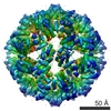
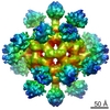
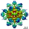
- EMDB-1558: Molecular Architecture of the 'stressosome', a signal transduction hub -
+ Open data

- Basic information
| Entry | Database: EMDB / ID: EMD-1558 | |||||||||
|---|---|---|---|---|---|---|---|---|---|---|
| Title | Molecular Architecture of the 'stressosome', a signal transduction hub | |||||||||
 Map data Map data | RsbR146-274RsbS Stressosome core with imposed D2 symmetry | |||||||||
 Sample Sample |
| |||||||||
 Keywords Keywords | RsbR / RsbS / Stressosome / sigmaB / RsbT / stress response / bacillus | |||||||||
| Biological species |  | |||||||||
| Method | single particle reconstruction / cryo EM / Resolution: 7.1 Å | |||||||||
 Authors Authors | Marles-Wright J / Grant T / Delumeau O / van Duinen G / Firbank SJ / Lewis PJ / Murray JW / Newman JA / Quin MB / Race PR ...Marles-Wright J / Grant T / Delumeau O / van Duinen G / Firbank SJ / Lewis PJ / Murray JW / Newman JA / Quin MB / Race PR / Rohou A / Tichelaar W / van Heel M / Lewis RJ | |||||||||
 Citation Citation | Journal: Science / Year: 2008 Title: Molecular architecture of the "stressosome," a signal integration and transduction hub. Authors: Jon Marles-Wright / Tim Grant / Olivier Delumeau / Gijs van Duinen / Susan J Firbank / Peter J Lewis / James W Murray / Joseph A Newman / Maureen B Quin / Paul R Race / Alexis Rohou / Willem ...Authors: Jon Marles-Wright / Tim Grant / Olivier Delumeau / Gijs van Duinen / Susan J Firbank / Peter J Lewis / James W Murray / Joseph A Newman / Maureen B Quin / Paul R Race / Alexis Rohou / Willem Tichelaar / Marin van Heel / Richard J Lewis /  Abstract: A commonly used strategy by microorganisms to survive multiple stresses involves a signal transduction cascade that increases the expression of stress-responsive genes. Stress signals can be ...A commonly used strategy by microorganisms to survive multiple stresses involves a signal transduction cascade that increases the expression of stress-responsive genes. Stress signals can be integrated by a multiprotein signaling hub that responds to various signals to effect a single outcome. We obtained a medium-resolution cryo-electron microscopy reconstruction of the 1.8-megadalton "stressosome" from Bacillus subtilis. Fitting known crystal structures of components into this reconstruction gave a pseudoatomic structure, which had a virus capsid-like core with sensory extensions. We suggest that the different sensory extensions respond to different signals, whereas the conserved domains in the core integrate the varied signals. The architecture of the stressosome provides the potential for cooperativity, suggesting that the response could be tuned dependent on the magnitude of chemophysical insult. | |||||||||
| History |
|
- Structure visualization
| Movie |
 Movie viewer Movie viewer |
|---|---|
| Structure viewer | EM map: SurfViewMolmilJmol/JSmol |
| Supplemental images |

- Downloads & links
-EMDB archive
| Map data | emd_1558.map.gz | 2.3 MB | EMDB map data format | |
|---|---|---|---|---|
| Header (meta data) | emd-1558-v30.xmlemd-1558.xml | 7.9 KB 7.9 KB | Display Display | EMDB header |
| Images |  emd1558.png emd1558.png | 275.7 KB | ||
| Archive directory |  http://ftp.pdbj.org/pub/emdb/structures/EMD-1558ftp://ftp.pdbj.org/pub/emdb/structures/EMD-1558 http://ftp.pdbj.org/pub/emdb/structures/EMD-1558ftp://ftp.pdbj.org/pub/emdb/structures/EMD-1558 | HTTPS FTP |
-Validation report
| Summary document | emd_1558_validation.pdf.gz | 243.3 KB | Display | EMDB validaton report |
|---|---|---|---|---|
| Full document | emd_1558_full_validation.pdf.gz | 242.4 KB | Display | |
| Data in XML | emd_1558_validation.xml.gz | 5.5 KB | Display | |
| Arichive directory | https://ftp.pdbj.org/pub/emdb/validation_reports/EMD-1558ftp://ftp.pdbj.org/pub/emdb/validation_reports/EMD-1558 | HTTPS FTP |
-Related structure data


-Links
| EMDB pages | EMDB (EBI/PDBe) / EMDataResource |
|---|
-Map
| File | Download / File: emd_1558.map.gz / Format: CCP4 / Size: 3.7 MB / Type: IMAGE STORED AS FLOATING POINT NUMBER (4 BYTES) | ||||||||||||||||||||||||||||||||||||||||||||||||||||||||||||||||||||
|---|---|---|---|---|---|---|---|---|---|---|---|---|---|---|---|---|---|---|---|---|---|---|---|---|---|---|---|---|---|---|---|---|---|---|---|---|---|---|---|---|---|---|---|---|---|---|---|---|---|---|---|---|---|---|---|---|---|---|---|---|---|---|---|---|---|---|---|---|---|
| Annotation | RsbR146-274RsbS Stressosome core with imposed D2 symmetry | ||||||||||||||||||||||||||||||||||||||||||||||||||||||||||||||||||||
| Voxel size | X=Y=Z: 2.54 Å | ||||||||||||||||||||||||||||||||||||||||||||||||||||||||||||||||||||
| Density |
| ||||||||||||||||||||||||||||||||||||||||||||||||||||||||||||||||||||
| Symmetry | Space group: 1 | ||||||||||||||||||||||||||||||||||||||||||||||||||||||||||||||||||||
| Details | EMDB XML:
CCP4 map header:
| ||||||||||||||||||||||||||||||||||||||||||||||||||||||||||||||||||||
-Supplemental data
- Sample components
-Entire : RsbR146-274RsbS stressosome core
| Entire | Name: RsbR146-274RsbS stressosome core |
|---|---|
| Components |
|
-Supramolecule #1000: RsbR146-274RsbS stressosome core
| Supramolecule | Name: RsbR146-274RsbS stressosome core / type: sample / ID: 1000 / Number unique components: 1 |
|---|
-Macromolecule #1: RsbR146-274RsbS Stressosome core
| Macromolecule | Name: RsbR146-274RsbS Stressosome core / type: protein_or_peptide / ID: 1 / Name.synonym: Stressosome core / Recombinant expression: Yes |
|---|---|
| Source (natural) | Organism: |
-Experimental details
-Structure determination
| Method | cryo EM |
|---|---|
 Processing Processing | single particle reconstruction |
| Aggregation state | particle |
-Sample preparation
| Vitrification | Cryogen name: ETHANE / Instrument: OTHER / Details: Vitrification instrument: Vitrobot |
|---|
- Electron microscopy
| Microscope | FEI/PHILIPS CM200FEG |
|---|---|
| Image recording | Digitization - Scanner: NIKON SUPER COOLSCAN 9000 |
| Electron beam | Acceleration voltage: 200 kV / Electron source:  FIELD EMISSION GUN FIELD EMISSION GUN |
| Electron optics | Illumination mode: FLOOD BEAM / Imaging mode: BRIGHT FIELD |
| Sample stage | Specimen holder: Side entry liquid nitrogen-cooled cryo specimen holder Specimen holder model: GATAN LIQUID NITROGEN |
-Image processing
| Final reconstruction | Applied symmetry - Point group: D2 (2x2 fold dihedral) / Resolution.type: BY AUTHOR / Resolution: 7.1 Å / Resolution method: OTHER / Software - Name: IMAGIC / Number images used: 13317 |
|---|
-Atomic model buiding 1
| Initial model | PDB ID: |
|---|---|
| Details | R and S were fitted using an homology model made using 2VY9 |
| Refinement | Space: REAL |
