 Movie
Movie Controller

[English] 日本語
 Yorodumi
- EMDB-1233: Structure of eEF3 and the mechanism of transfer RNA release from ... -
+ Open data

- Basic information
| Entry | Database: EMDB / ID: EMD-1233 | |||||||||
|---|---|---|---|---|---|---|---|---|---|---|
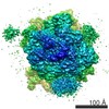
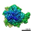
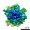
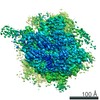
| Title | Structure of eEF3 and the mechanism of transfer RNA release from the E-site. | |||||||||
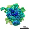
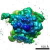
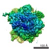
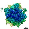
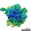
 Map data Map data | Cryo-EM density map of yeast eEF3 bound to translating yeast 80S ribosome | |||||||||
 Sample Sample |
| |||||||||
| Function / homology |  Function and homology information Function and homology informationtranslational elongation / translation elongation factor activity / translational termination / cytosolic ribosome / negative regulation of protein phosphorylation / negative regulation of protein kinase activity / Hydrolases; Acting on acid anhydrides; Acting on acid anhydrides to facilitate cellular and subcellular movement / cytoplasmic stress granule / ribosome binding / rRNA binding ...translational elongation / translation elongation factor activity / translational termination / cytosolic ribosome / negative regulation of protein phosphorylation / negative regulation of protein kinase activity / Hydrolases; Acting on acid anhydrides; Acting on acid anhydrides to facilitate cellular and subcellular movement / cytoplasmic stress granule / ribosome binding / rRNA binding / ribosome / ATP hydrolysis activity / ATP binding Similarity search - Function | |||||||||
| Biological species |  | |||||||||
| Method | single particle reconstruction / cryo EM / negative staining / Resolution: 9.9 Å | |||||||||
 Authors Authors | Beckmann R / Andersen G | |||||||||
 Citation Citation | Journal: Nature / Year: 2006 Title: Structure of eEF3 and the mechanism of transfer RNA release from the E-site. Authors: Christian B F Andersen / Thomas Becker / Michael Blau / Monika Anand / Mario Halic / Bharvi Balar / Thorsten Mielke / Thomas Boesen / Jan Skov Pedersen / Christian M T Spahn / Terri Goss ...Authors: Christian B F Andersen / Thomas Becker / Michael Blau / Monika Anand / Mario Halic / Bharvi Balar / Thorsten Mielke / Thomas Boesen / Jan Skov Pedersen / Christian M T Spahn / Terri Goss Kinzy / Gregers R Andersen / Roland Beckmann /  Abstract: Elongation factor eEF3 is an ATPase that, in addition to the two canonical factors eEF1A and eEF2, serves an essential function in the translation cycle of fungi. eEF3 is required for the binding of ...Elongation factor eEF3 is an ATPase that, in addition to the two canonical factors eEF1A and eEF2, serves an essential function in the translation cycle of fungi. eEF3 is required for the binding of the aminoacyl-tRNA-eEF1A-GTP ternary complex to the ribosomal A-site and has been suggested to facilitate the clearance of deacyl-tRNA from the E-site. Here we present the crystal structure of Saccharomyces cerevisiae eEF3, showing that it consists of an amino-terminal HEAT repeat domain, followed by a four-helix bundle and two ABC-type ATPase domains, with a chromodomain inserted in ABC2. Moreover, we present the cryo-electron microscopy structure of the ATP-bound form of eEF3 in complex with the post-translocational-state 80S ribosome from yeast. eEF3 uses an entirely new factor binding site near the ribosomal E-site, with the chromodomain likely to stabilize the ribosomal L1 stalk in an open conformation, thus allowing tRNA release. | |||||||||
| History |
|
- Structure visualization
| Movie |
Movie viewer |
|---|---|
| Structure viewer | EM map: SurfViewMolmilJmol/JSmol |
| Supplemental images |

- Downloads & links
-EMDB archive
| Map data | emd_1233.map.gz | 73.8 MB | EMDB map data format | |
|---|---|---|---|---|
| Header (meta data) | emd-1233-v30.xmlemd-1233.xml | 11.7 KB 11.7 KB | Display Display | EMDB header |
| Images |  1233.gif 1233.gif | 18.5 KB | ||
| Archive directory |  http://ftp.pdbj.org/pub/emdb/structures/EMD-1233ftp://ftp.pdbj.org/pub/emdb/structures/EMD-1233 http://ftp.pdbj.org/pub/emdb/structures/EMD-1233ftp://ftp.pdbj.org/pub/emdb/structures/EMD-1233 | HTTPS FTP |
-Validation report
| Summary document | emd_1233_validation.pdf.gz | 286.9 KB | Display | EMDB validaton report |
|---|---|---|---|---|
| Full document | emd_1233_full_validation.pdf.gz | 286 KB | Display | |
| Data in XML | emd_1233_validation.xml.gz | 5.5 KB | Display | |
| Arichive directory | https://ftp.pdbj.org/pub/emdb/validation_reports/EMD-1233ftp://ftp.pdbj.org/pub/emdb/validation_reports/EMD-1233 | HTTPS FTP |
-Related structure data
| Related structure data | 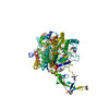 2ix8MC 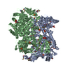 2iw3C 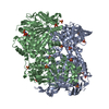 2iwhC 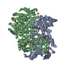 2ix3C M: atomic model generated by this map C: citing same article ( |
|---|---|
| Similar structure data |

-Links
| EMDB pages | EMDB (EBI/PDBe) / EMDataResource |
|---|---|
| Related items in Molecule of the Month |


-Map
| File | Download / File: emd_1233.map.gz / Format: CCP4 / Size: 78.3 MB / Type: IMAGE STORED AS FLOATING POINT NUMBER (4 BYTES) | ||||||||||||||||||||||||||||||||||||||||||||||||||||||||||||||||||||
|---|---|---|---|---|---|---|---|---|---|---|---|---|---|---|---|---|---|---|---|---|---|---|---|---|---|---|---|---|---|---|---|---|---|---|---|---|---|---|---|---|---|---|---|---|---|---|---|---|---|---|---|---|---|---|---|---|---|---|---|---|---|---|---|---|---|---|---|---|---|
| Annotation | Cryo-EM density map of yeast eEF3 bound to translating yeast 80S ribosome | ||||||||||||||||||||||||||||||||||||||||||||||||||||||||||||||||||||
| Voxel size |
| ||||||||||||||||||||||||||||||||||||||||||||||||||||||||||||||||||||
| Density |
| ||||||||||||||||||||||||||||||||||||||||||||||||||||||||||||||||||||
| Symmetry | Space group: 1 | ||||||||||||||||||||||||||||||||||||||||||||||||||||||||||||||||||||
| Details | EMDB XML:
CCP4 map header:
| ||||||||||||||||||||||||||||||||||||||||||||||||||||||||||||||||||||
-Supplemental data
- Sample components
-Entire : 80S-RNC-eEF3-AMP-PNP complex from S. cerevisiae
| Entire | Name: 80S-RNC-eEF3-AMP-PNP complex from S. cerevisiae |
|---|---|
| Components |
|
-Supramolecule #1000: 80S-RNC-eEF3-AMP-PNP complex from S. cerevisiae
| Supramolecule | Name: 80S-RNC-eEF3-AMP-PNP complex from S. cerevisiae / type: sample / ID: 1000 Details: The emerging signal sequence of the ribosome nascent chain (RNC) was saturated using purified trimeric Sec61 complex in order to prevent biased orientation of particles Oligomeric state: One ribosome binds to one molecule of eEF3 Number unique components: 2 |
|---|---|
| Molecular weight | Theoretical: 4.3 MDa |
-Supramolecule #1: programmed 80S ribosome
| Supramolecule | Name: programmed 80S ribosome / type: complex / ID: 1 / Name.synonym: RNC / Details: programmed 80S ribosome with a P-site tRNA / Ribosome-details: ribosome-eukaryote: ALL |
|---|---|
| Molecular weight | Experimental: 4.2 MDa / Theoretical: 4.2 MDa |
-Macromolecule #1: elongation factor 3
| Macromolecule | Name: elongation factor 3 / type: protein_or_peptide / ID: 1 / Name.synonym: eEF3 / Details: eEF3 with a C-terminal His-Tag and a factor / Number of copies: 1 / Oligomeric state: Monomer / Recombinant expression: Yes |
|---|---|
| Source (natural) | Organism: |
| Molecular weight | Experimental: 115 KDa / Theoretical: 115 KDa |
| Recombinant expression | Organism: |
-Experimental details
-Structure determination
| Method | negative staining, cryo EM |
|---|---|
 Processing Processing | single particle reconstruction |
| Aggregation state | particle |
-Sample preparation
| Concentration | 0.168 mg/mL |
|---|---|
| Buffer | pH: 7.5 Details: 20 mM HEPES, pH 7.5, 10 mM Mg(OAc)2, 150 mM KOAc 1 mM DTT, 0.05% Nikkol, 125 mM Sucrose,0.01 mg ml-1 Cycloheximide, 0.5 mM AMP-PNP, 0.1 mM Neomycin, 0.3 % DesoxyBigChaps |
| Staining | Type: NEGATIVE / Details: Cryo-EM, no staining |
| Grid | Details: Quantifoil grids 200 mesh R2/4 |
| Vitrification | Cryogen name: ETHANE / Chamber humidity: 95 % / Instrument: OTHER / Details: Vitrification instrument: Vitrobot Method: Blot for 10 seconds before plunging, use 2 layers of filter paper |
- Electron microscopy
| Microscope | FEI TECNAI F30 |
|---|---|
| Temperature | Min: 95 K / Average: 95 K |
| Alignment procedure | Legacy - Astigmatism: objective lens astigmatism was corrected at 100,000 time |
| Date | Jan 13, 2005 |
| Image recording | Category: FILM / Film or detector model: KODAK SO-163 FILM / Digitization - Scanner: PRIMESCAN / Digitization - Sampling interval: 4.35 µm / Number real images: 141 / Average electron dose: 20 e/Å2 / Od range: 1.2 / Bits/pixel: 16 |
| Electron beam | Acceleration voltage: 300 kV / Electron source:  FIELD EMISSION GUN FIELD EMISSION GUN |
| Electron optics | Calibrated magnification: 38900 / Illumination mode: SPOT SCAN / Imaging mode: BRIGHT FIELD / Cs: 2.27 mm / Nominal defocus max: 3.5 µm / Nominal defocus min: 1.3 µm / Nominal magnification: 39000 |
| Sample stage | Specimen holder: Polara Multispecimen Holder / Specimen holder model: OTHER |
| Experimental equipment |  Model: Tecnai F30 / Image courtesy: FEI Company |
-Image processing
| Final reconstruction | Applied symmetry - Point group: C1 (asymmetric) / Algorithm: OTHER / Resolution.type: BY AUTHOR / Resolution: 9.9 Å / Resolution method: FSC 0.5 CUT-OFF / Software - Name: SPIDER / Number images used: 37700 |
|---|
-Atomic model buiding 1
| Software | Name: O and Situs |
|---|---|
| Details | Protocol: rigid body. The domains were separately fitted by manual docking using program O |
| Refinement | Protocol: RIGID BODY FIT / Target criteria: cross correlation |
| Output model | PDB-2ix8: |
