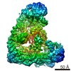
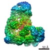
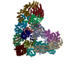
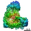
Journal: Proc Natl Acad Sci U S A / Year: 2019 Title: Protein engineering of a ubiquitin-variant inhibitor of APC/C identifies a cryptic K48 ubiquitin chain binding site. Authors: Edmond R Watson / Christy R R Grace / Wei Zhang / Darcie J Miller / Iain F Davidson / J Rajan Prabu / Shanshan Yu / Derek L Bolhuis / Elizaveta T Kulko / Ronnald Vollrath / David Haselbach / ...Authors: Edmond R Watson / Christy R R Grace / Wei Zhang / Darcie J Miller / Iain F Davidson / J Rajan Prabu / Shanshan Yu / Derek L Bolhuis / Elizaveta T Kulko / Ronnald Vollrath / David Haselbach / Holger Stark / Jan-Michael Peters / Nicholas G Brown / Sachdev S Sidhu / Brenda A Schulman / Abstract: Ubiquitin (Ub)-mediated proteolysis is a fundamental mechanism used by eukaryotic cells to maintain homeostasis and protein quality, and to control timing in biological processes. Two essential ...Ubiquitin (Ub)-mediated proteolysis is a fundamental mechanism used by eukaryotic cells to maintain homeostasis and protein quality, and to control timing in biological processes. Two essential aspects of Ub regulation are conjugation through E1-E2-E3 enzymatic cascades and recognition by Ub-binding domains. An emerging theme in the Ub field is that these 2 properties are often amalgamated in conjugation enzymes. In addition to covalent thioester linkage to Ub's C terminus for Ub transfer reactions, conjugation enzymes often bind noncovalently and weakly to Ub at "exosites." However, identification of such sites is typically empirical and particularly challenging in large molecular machines. Here, studying the 1.2-MDa E3 ligase anaphase-promoting complex/cyclosome (APC/C), which controls cell division and many aspects of neurobiology, we discover a method for identifying unexpected Ub-binding sites. Using a panel of Ub variants (UbVs), we identify a protein-based inhibitor that blocks Ub ligation to APC/C substrates in vitro and ex vivo. Biochemistry, NMR, and cryo-electron microscopy (cryo-EM) structurally define the UbV interaction, explain its inhibitory activity through binding the surface on the APC2 subunit that recruits the E2 enzyme UBE2C, and ultimately reveal that this APC2 surface is also a Ub-binding exosite with preference for K48-linked chains. The results provide a tool for probing APC/C activity, have implications for the coordination of K48-linked Ub chain binding by APC/C with the multistep process of substrate polyubiquitylation, and demonstrate the power of UbV technology for identifying cryptic Ub-binding sites within large multiprotein complexes.
History
Deposition
Jun 14, 2019
-
Header (metadata) release
Aug 7, 2019
-
Map release
Aug 7, 2019
-
Update
Nov 25, 2020
-
Current status
Nov 25, 2020
Processing site: PDBe / Status: Released
-
Structure visualization
Movie
Surface view with section colored by density value
In the structure databanks used in Yorodumi, some data are registered as the other names, "COVID-19 virus" and "2019-nCoV". Here are the details of the virus and the list of structure data.
Jan 31, 2019. EMDB accession codes are about to change! (news from PDBe EMDB page)
EMDB accession codes are about to change! (news from PDBe EMDB page)
The allocation of 4 digits for EMDB accession codes will soon come to an end. Whilst these codes will remain in use, new EMDB accession codes will include an additional digit and will expand incrementally as the available range of codes is exhausted. The current 4-digit format prefixed with “EMD-” (i.e. EMD-XXXX) will advance to a 5-digit format (i.e. EMD-XXXXX), and so on. It is currently estimated that the 4-digit codes will be depleted around Spring 2019, at which point the 5-digit format will come into force.
The EM Navigator/Yorodumi systems omit the EMD- prefix.
Related info.:Q: What is EMD? / ID/Accession-code notation in Yorodumi/EM Navigator
Yorodumi is a browser for structure data from EMDB, PDB, SASBDB, etc.
This page is also the successor to EM Navigator detail page, and also detail information page/front-end page for Omokage search.
The word "yorodu" (or yorozu) is an old Japanese word meaning "ten thousand". "mi" (miru) is to see.
Related info.:EMDB / PDB / SASBDB / Comparison of 3 databanks / Yorodumi Search / Aug 31, 2016. New EM Navigator & Yorodumi / Yorodumi Papers / Jmol/JSmol / Function and homology information / Changes in new EM Navigator and Yorodumi
 Movie
Movie Controller


 Open data
 Basic information
 Map data
 Sample
 Homo sapiens (human)
 Authors
 Germany, 2 items
  Citation


 Structure visualization
 Movie viewer
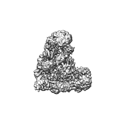
 Downloads & links
 emd_10072.png
 http://ftp.pdbj.org/pub/emdb/structures/EMD-10072


 Z
Z Y
Y X
X


 Sample components
 Processing
 Electron microscopy
