 Movie
Movie Controller
 Structure viewers
 About Yorodumi Papers


+Search query
-Structure paper
| Title | Protein engineering of a ubiquitin-variant inhibitor of APC/C identifies a cryptic K48 ubiquitin chain binding site. |
|---|---|
| Journal, issue, pages | Proc Natl Acad Sci U S A, Vol. 116, Issue 35, Page 17280-17289, Year 2019 |
| Publish date | Aug 27, 2019 |
 Authors Authors | Edmond R Watson / Christy R R Grace / Wei Zhang / Darcie J Miller / Iain F Davidson / J Rajan Prabu / Shanshan Yu / Derek L Bolhuis / Elizaveta T Kulko / Ronnald Vollrath / David Haselbach / Holger Stark / Jan-Michael Peters / Nicholas G Brown / Sachdev S Sidhu / Brenda A Schulman /     |
| PubMed Abstract | Ubiquitin (Ub)-mediated proteolysis is a fundamental mechanism used by eukaryotic cells to maintain homeostasis and protein quality, and to control timing in biological processes. Two essential ...Ubiquitin (Ub)-mediated proteolysis is a fundamental mechanism used by eukaryotic cells to maintain homeostasis and protein quality, and to control timing in biological processes. Two essential aspects of Ub regulation are conjugation through E1-E2-E3 enzymatic cascades and recognition by Ub-binding domains. An emerging theme in the Ub field is that these 2 properties are often amalgamated in conjugation enzymes. In addition to covalent thioester linkage to Ub's C terminus for Ub transfer reactions, conjugation enzymes often bind noncovalently and weakly to Ub at "exosites." However, identification of such sites is typically empirical and particularly challenging in large molecular machines. Here, studying the 1.2-MDa E3 ligase anaphase-promoting complex/cyclosome (APC/C), which controls cell division and many aspects of neurobiology, we discover a method for identifying unexpected Ub-binding sites. Using a panel of Ub variants (UbVs), we identify a protein-based inhibitor that blocks Ub ligation to APC/C substrates in vitro and ex vivo. Biochemistry, NMR, and cryo-electron microscopy (cryo-EM) structurally define the UbV interaction, explain its inhibitory activity through binding the surface on the APC2 subunit that recruits the E2 enzyme UBE2C, and ultimately reveal that this APC2 surface is also a Ub-binding exosite with preference for K48-linked chains. The results provide a tool for probing APC/C activity, have implications for the coordination of K48-linked Ub chain binding by APC/C with the multistep process of substrate polyubiquitylation, and demonstrate the power of UbV technology for identifying cryptic Ub-binding sites within large multiprotein complexes. |
 External links External links | Proc Natl Acad Sci U S A / PubMed:31350353 / PubMed Central |
| Methods | EM (single particle) / X-ray diffraction / NMR (solution) |
| Resolution | 2.2 - 6.6 Å |
| Structure data | 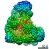 EMDB-10072:  PDB-6nxk: 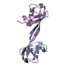 PDB-6nxl:  PDB-6ob1: |
| Chemicals |  ChemComp-HOH: |
| Source |
|
 Keywords Keywords |  LIGASE / APC / WHB / Ubiquitin / Diubiquitin / Ubv / Inhibitor / PROTEIN BINDING LIGASE / APC / WHB / Ubiquitin / Diubiquitin / Ubv / Inhibitor / PROTEIN BINDING |

