 Movie
Movie Controller


+ Open data

- Basic information
| Entry | Database: PDB / ID: 8h1o | |||||||||||||||||||||||||||||||||||||||||||||||||||
|---|---|---|---|---|---|---|---|---|---|---|---|---|---|---|---|---|---|---|---|---|---|---|---|---|---|---|---|---|---|---|---|---|---|---|---|---|---|---|---|---|---|---|---|---|---|---|---|---|---|---|---|---|
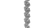
| Title | Cryo-EM structure of KpFtsZ-monobody double helical tube | |||||||||||||||||||||||||||||||||||||||||||||||||||
 Components Components |
| |||||||||||||||||||||||||||||||||||||||||||||||||||
 Keywords Keywords |  CELL CYCLE / bacterial cell division / divisome / FtsZ / monobody / tubulin CELL CYCLE / bacterial cell division / divisome / FtsZ / monobody / tubulin | |||||||||||||||||||||||||||||||||||||||||||||||||||
| Function / homology |  Function and homology information Function and homology informationFtsZ-dependent cytokinesis / division septum assembly / cell division site / protein polymerization / GTPase activity / GTP binding / cytoplasmSimilarity search - Function | |||||||||||||||||||||||||||||||||||||||||||||||||||
| Biological species |  Klebsiella pneumoniae (bacteria) Klebsiella pneumoniae (bacteria) Homo sapiens (human) Homo sapiens (human) | |||||||||||||||||||||||||||||||||||||||||||||||||||
| Method | ELECTRON MICROSCOPY / helical reconstruction / cryo EM / Resolution: 2.67 Å | |||||||||||||||||||||||||||||||||||||||||||||||||||
 Authors Authors | Fujita, J. / Amesaka, H. / Yoshizawa, T. / Kuroda, N. / Kamimura, N. / Hara, M. / Inoue, T. / Namba, K. / Tanaka, S. / Matsumura, H. | |||||||||||||||||||||||||||||||||||||||||||||||||||
| Funding support |  Japan, 16items Japan, 16items
| |||||||||||||||||||||||||||||||||||||||||||||||||||
 Citation Citation | Journal: Nat Commun / Year: 2023 Title: Structures of a FtsZ single protofilament and a double-helical tube in complex with a monobody. Authors: Junso Fujita / Hiroshi Amesaka / Takuya Yoshizawa / Kota Hibino / Natsuki Kamimura / Natsuko Kuroda / Takamoto Konishi / Yuki Kato / Mizuho Hara / Tsuyoshi Inoue / Keiichi Namba / Shun-Ichi ...Authors: Junso Fujita / Hiroshi Amesaka / Takuya Yoshizawa / Kota Hibino / Natsuki Kamimura / Natsuko Kuroda / Takamoto Konishi / Yuki Kato / Mizuho Hara / Tsuyoshi Inoue / Keiichi Namba / Shun-Ichi Tanaka / Hiroyoshi Matsumura / Abstract: FtsZ polymerizes into protofilaments to form the Z-ring that acts as a scaffold for accessory proteins during cell division. Structures of FtsZ have been previously solved, but detailed mechanistic ...FtsZ polymerizes into protofilaments to form the Z-ring that acts as a scaffold for accessory proteins during cell division. Structures of FtsZ have been previously solved, but detailed mechanistic insights are lacking. Here, we determine the cryoEM structure of a single protofilament of FtsZ from Klebsiella pneumoniae (KpFtsZ) in a polymerization-preferred conformation. We also develop a monobody (Mb) that binds to KpFtsZ and FtsZ from Escherichia coli without affecting their GTPase activity. Crystal structures of the FtsZ-Mb complexes reveal the Mb binding mode, while addition of Mb in vivo inhibits cell division. A cryoEM structure of a double-helical tube of KpFtsZ-Mb at 2.7 Å resolution shows two parallel protofilaments. Our present study highlights the physiological roles of the conformational changes of FtsZ in treadmilling that regulate cell division. | |||||||||||||||||||||||||||||||||||||||||||||||||||
| History |
|
- Structure visualization
| Structure viewer | Molecule: MolmilJmol/JSmol |
|---|
- Downloads & links
-Download
| PDBx/mmCIF format | 8h1o.cif.gz | 82.9 KB | Display | PDBx/mmCIF format |
|---|---|---|---|---|
| PDB format | pdb8h1o.ent.gz | 60.8 KB | Display | PDB format |
| PDBx/mmJSON format | 8h1o.json.gz | Tree view | PDBx/mmJSON format | |
| Others |  Other downloads Other downloads |
-Validation report
| Arichive directory | https://data.pdbj.org/pub/pdb/validation_reports/h1/8h1oftp://data.pdbj.org/pub/pdb/validation_reports/h1/8h1o | HTTPS FTP |
|---|
-Related structure data
| Related structure data | 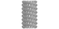 34429MC 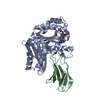 8gzvC  8gzwC  8gzxC  8gzyC 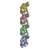 8ibnC C: citing same article ( M: map data used to model this data |
|---|---|
| Similar structure data |
-Links
 PDBj
PDBj


- Assembly
| Deposited unit | 
|
|---|---|
| 1 | x 100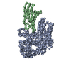
|
-Components
| #1: Protein | Mass: 40574.926 Da / Num. of mol.: 1 Source method: isolated from a genetically manipulated source Source: (gene. exp.) Klebsiella pneumoniae (bacteria) / Production host: Escherichia coli (E. coli) / References: UniProt: W9BCK7 |
|---|---|
| #2: Protein | Mass: 9781.873 Da / Num. of mol.: 1 Source method: isolated from a genetically manipulated source Source: (gene. exp.) Homo sapiens (human) / Production host: Escherichia coli (E. coli) |
| #3: Chemical | ChemComp-GDP / Guanosine diphosphate  Type: RNA linking / Mass: 443.201 Da / Num. of mol.: 1 / Source method: obtained synthetically / Formula: C10H15N5O11P2 / Feature type: SUBJECT OF INVESTIGATION / Comment: GDP, energy-carrying molecule*YM Type: RNA linking / Mass: 443.201 Da / Num. of mol.: 1 / Source method: obtained synthetically / Formula: C10H15N5O11P2 / Feature type: SUBJECT OF INVESTIGATION / Comment: GDP, energy-carrying molecule*YM |
| Has ligand of interest | Y |
-Experimental details
-Experiment
| Experiment | Method: ELECTRON MICROSCOPY |
|---|---|
| EM experiment | Aggregation state: FILAMENT / 3D reconstruction method: helical reconstruction |
- Sample preparation
| Component |
| |||||||||||||||||||||||||
|---|---|---|---|---|---|---|---|---|---|---|---|---|---|---|---|---|---|---|---|---|---|---|---|---|---|---|
| Molecular weight | Experimental value: NO | |||||||||||||||||||||||||
| Source (natural) |
| |||||||||||||||||||||||||
| Source (recombinant) |
| |||||||||||||||||||||||||
| Buffer solution | pH: 7.5 Details: 1 mM GMPPNP and 0.12 mM PC190723 were supplemented. | |||||||||||||||||||||||||
| Buffer component |
| |||||||||||||||||||||||||
| Specimen | Conc.: 4 mg/ml / Embedding applied: NO / Shadowing applied: NO / Staining applied: NO / Vitrification applied: YES / Details: 1.2x molar excess of Mb was supplemented. | |||||||||||||||||||||||||
| Specimen support | Details: The graphene grid was chemically oxidized and modified. Grid material: GOLD / Grid mesh size: 200 divisions/in. / Grid type: Quantifoil R1.2/1.3 | |||||||||||||||||||||||||
| Vitrification | Instrument: FEI VITROBOT MARK IV / Cryogen name: ETHANE |
- Electron microscopy imaging
| Microscopy | Model: JEOL CRYO ARM 300 |
|---|---|
| Electron gun | Electron source: FIELD EMISSION GUN / Accelerating voltage: 300 kV / Illumination mode: FLOOD BEAM |
| Electron lens | Mode: BRIGHT FIELDBright-field microscopy / Nominal magnification: 60000 X / Nominal defocus max: 2000 nm / Nominal defocus min: 500 nm / Cs: 2.7 mm |
| Specimen holder | Cryogen: NITROGEN / Specimen holder model: JEOL CRYOSPECPORTER |
| Image recording | Average exposure time: 3 sec. / Electron dose: 60 e/Å2 / Film or detector model: GATAN K3 (6k x 4k) / Num. of grids imaged: 1 |
| EM imaging optics | Energyfilter name: In-column Omega Filter / Energyfilter slit width: 20 eV |
- Processing
| Software |
| ||||||||||||||||||||||||||||||||||||
|---|---|---|---|---|---|---|---|---|---|---|---|---|---|---|---|---|---|---|---|---|---|---|---|---|---|---|---|---|---|---|---|---|---|---|---|---|---|
| EM software |
| ||||||||||||||||||||||||||||||||||||
| CTF correction | Type: PHASE FLIPPING AND AMPLITUDE CORRECTION | ||||||||||||||||||||||||||||||||||||
| Helical symmerty | Angular rotation/subunit: -23.398 ° / Axial rise/subunit: 7.703 Å / Axial symmetry: C2 | ||||||||||||||||||||||||||||||||||||
| Particle selection | Num. of particles selected: 181247 | ||||||||||||||||||||||||||||||||||||
| 3D reconstruction | Resolution: 2.67 Å / Resolution method: FSC 0.143 CUT-OFF / Num. of particles: 90711 / Algorithm: FOURIER SPACE / Symmetry type: HELICAL | ||||||||||||||||||||||||||||||||||||
| Atomic model building | Space: REAL | ||||||||||||||||||||||||||||||||||||
| Refinement | Cross valid method: NONE Stereochemistry target values: GeoStd + Monomer Library + CDL v1.2 | ||||||||||||||||||||||||||||||||||||
| Displacement parameters | Biso mean: 34.49 Å2 | ||||||||||||||||||||||||||||||||||||
| Refine LS restraints |
|
