 Movie
Movie Controller


+ Open data

- Basic information
| Entry |  | |||||||||||||||||||||||||||||||||||||||||||||||||||
|---|---|---|---|---|---|---|---|---|---|---|---|---|---|---|---|---|---|---|---|---|---|---|---|---|---|---|---|---|---|---|---|---|---|---|---|---|---|---|---|---|---|---|---|---|---|---|---|---|---|---|---|---|
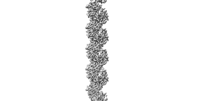
| Title | Cryo-EM structure of KpFtsZ single filament | |||||||||||||||||||||||||||||||||||||||||||||||||||
 Map data Map data | ||||||||||||||||||||||||||||||||||||||||||||||||||||
 Sample Sample |
| |||||||||||||||||||||||||||||||||||||||||||||||||||
 Keywords Keywords | bacterial cell division /  divisome / monobody / CELL CYCLE divisome / monobody / CELL CYCLE | |||||||||||||||||||||||||||||||||||||||||||||||||||
| Function / homology |  Function and homology information Function and homology informationFtsZ-dependent cytokinesis / division septum assembly / cell division site / protein polymerization / GTPase activity / GTP binding / cytoplasmSimilarity search - Function | |||||||||||||||||||||||||||||||||||||||||||||||||||
| Biological species |  Klebsiella pneumoniae (bacteria) Klebsiella pneumoniae (bacteria) | |||||||||||||||||||||||||||||||||||||||||||||||||||
| Method | helical reconstruction / cryo EM / Resolution: 3.03 Å | |||||||||||||||||||||||||||||||||||||||||||||||||||
 Authors Authors | Fujita J / Amesaka H / Yoshizawa T / Kuroda N / Kamimura N / Hibino K / Konishi T / Kato Y / Hara M / Inoue T ...Fujita J / Amesaka H / Yoshizawa T / Kuroda N / Kamimura N / Hibino K / Konishi T / Kato Y / Hara M / Inoue T / Namba K / Tanaka S / Matsumura H | |||||||||||||||||||||||||||||||||||||||||||||||||||
| Funding support |  Japan, 16 items Japan, 16 items
| |||||||||||||||||||||||||||||||||||||||||||||||||||
 Citation Citation | Journal: Nat Commun / Year: 2023 Title: Structures of a FtsZ single protofilament and a double-helical tube in complex with a monobody Authors: Fujita J / Amesaka H / Yoshizawa T / Hibino K / Kamimura N / Kuroda N / Konishi T / Kato Y / Hara M / Inoue T / Namba K / Tanaka SI / Matsumura H | |||||||||||||||||||||||||||||||||||||||||||||||||||
| History |
|
- Structure visualization
| Supplemental images |
|---|
- Downloads & links
-EMDB archive
| Map data | emd_35344.map.gz | 97.3 MB | EMDB map data format | |
|---|---|---|---|---|
| Header (meta data) | emd-35344-v30.xmlemd-35344.xml | 22 KB 22 KB | Display Display | EMDB header |
| FSC (resolution estimation) | emd_35344_fsc.xml | 9.9 KB | Display | FSC data file |
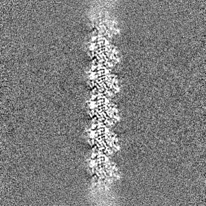
| Images |  emd_35344.png emd_35344.png | 22.7 KB | ||
| Masks | emd_35344_msk_1.map | 103 MB | Mask map | |
| Others | emd_35344_half_map_1.map.gzemd_35344_half_map_2.map.gz | 95.5 MB 95.5 MB | ||
| Archive directory |  http://ftp.pdbj.org/pub/emdb/structures/EMD-35344ftp://ftp.pdbj.org/pub/emdb/structures/EMD-35344 http://ftp.pdbj.org/pub/emdb/structures/EMD-35344ftp://ftp.pdbj.org/pub/emdb/structures/EMD-35344 | HTTPS FTP |
-Related structure data
| Related structure data | 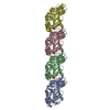 8ibnMC M: atomic model generated by this map C: citing same article ( |
|---|---|
| Similar structure data |
-Links
| EMDB pages | EMDB (EBI/PDBe) / EMDataResource |
|---|---|
| Related items in Molecule of the Month |


-Map
| File | Download / File: emd_35344.map.gz / Format: CCP4 / Size: 103 MB / Type: IMAGE STORED AS FLOATING POINT NUMBER (4 BYTES) | ||||||||||||||||||||
|---|---|---|---|---|---|---|---|---|---|---|---|---|---|---|---|---|---|---|---|---|---|
| Voxel size | X=Y=Z: 1.0344 Å | ||||||||||||||||||||
| Density |
| ||||||||||||||||||||
| Symmetry | Space group: 1 | ||||||||||||||||||||
| Details | EMDB XML:
|
-Supplemental data
-Mask #1
| File | emd_35344_msk_1.map | ||||||||||||
|---|---|---|---|---|---|---|---|---|---|---|---|---|---|
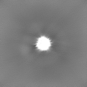
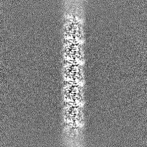
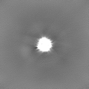
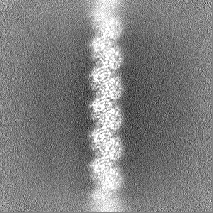
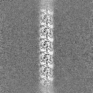
| Projections & Slices |
| ||||||||||||
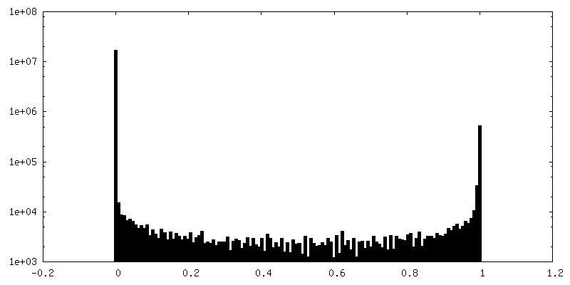
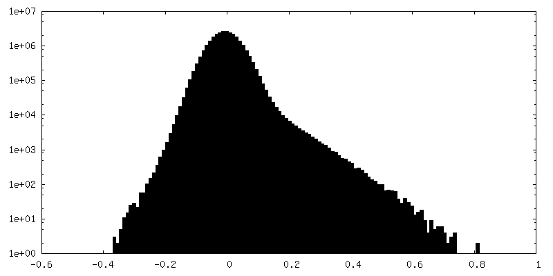
| Density Histograms |
 Z
Z Y
Y X
X


-Half map: #2
| File | emd_35344_half_map_1.map | ||||||||||||
|---|---|---|---|---|---|---|---|---|---|---|---|---|---|
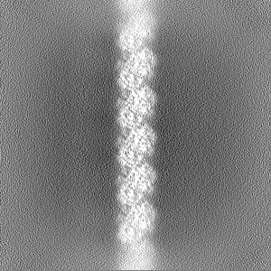
| Projections & Slices |
| ||||||||||||
| Density Histograms |


-Half map: #1
| File | emd_35344_half_map_2.map | ||||||||||||
|---|---|---|---|---|---|---|---|---|---|---|---|---|---|
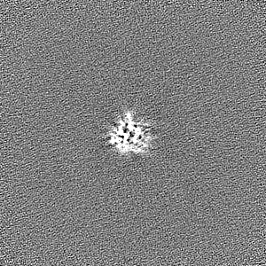
| Projections & Slices |
| ||||||||||||
| Density Histograms |


- Sample components
-Entire : Cryo-EM structure of KpFtsZ single filament
| Entire | Name: Cryo-EM structure of KpFtsZ single filament |
|---|---|
| Components |
|
-Supramolecule #1: Cryo-EM structure of KpFtsZ single filament
| Supramolecule | Name: Cryo-EM structure of KpFtsZ single filament / type: complex / ID: 1 / Parent: 0 / Macromolecule list: #1 |
|---|
-Supramolecule #2: KpFtsZ
| Supramolecule | Name: KpFtsZ / type: complex / ID: 2 / Parent: 1 / Macromolecule list: #1 |
|---|---|
| Source (natural) | Organism: Klebsiella pneumoniae (bacteria) |
-Macromolecule #1: Cell division protein FtsZ
| Macromolecule | Name: Cell division protein FtsZ / type: protein_or_peptide / ID: 1 / Number of copies: 4 / Enantiomer: LEVO |
|---|---|
| Source (natural) | Organism: Klebsiella pneumoniae (bacteria) |
| Molecular weight | Theoretical: 40.574926 KDa |
| Recombinant expression | Organism: Escherichia coli (E. coli) |
| Sequence | String: GHMFEPMELT NDAVIKVIGV GGGGGNAVEH MVRERIEGVE FFAVNTDAQA LRKTAVGQTI QIGSGITKGL GAGANPEVGR NAADEDREA LRAALDGADM VFIAAGMGGG TGTGAAPVVA EVAKDLGILT VAVVTKPFNF EGKKRMAFAE QGITELSKHV D SLITIPND ...String: GHMFEPMELT NDAVIKVIGV GGGGGNAVEH MVRERIEGVE FFAVNTDAQA LRKTAVGQTI QIGSGITKGL GAGANPEVGR NAADEDREA LRAALDGADM VFIAAGMGGG TGTGAAPVVA EVAKDLGILT VAVVTKPFNF EGKKRMAFAE QGITELSKHV D SLITIPND KLLKVLGRGI SLLDAFGAAN DVLKGAVQGI AELITRPGLM NVDFADVRTV MSEMGYAMMG SGVASGEDRA EE AAEMAIS SPLLEDIDLS GARGVLVNIT AGFDLRLDEF ETVGNTIRAF ASDNATVVIG TSLDPDMNDE LRVTVVATGI GMD KRPEIT LVTNKQVQQP VMDRYQQHGM SPLTQEQKPA AKVVNDNTPQ TAKEPDYLDI PAFLRKQAD UniProtKB: Cell division protein FtsZ |
-Macromolecule #2: PHOSPHOMETHYLPHOSPHONIC ACID GUANYLATE ESTER
| Macromolecule | Name: PHOSPHOMETHYLPHOSPHONIC ACID GUANYLATE ESTER / type: ligand / ID: 2 / Number of copies: 4 / Formula: G2P |
|---|---|
| Molecular weight | Theoretical: 521.208 Da |
| Chemical component information |  ChemComp-G2P: |
-Macromolecule #3: POTASSIUM ION
| Macromolecule | Name: POTASSIUM ION / type: ligand / ID: 3 / Number of copies: 4 / Formula: K |
|---|---|
| Molecular weight | Theoretical: 39.098 Da |
-Experimental details
-Structure determination
| Method | cryo EM |
|---|---|
 Processing Processing | helical reconstruction |
| Aggregation state | filament |
-Sample preparation
| Concentration | 0.5 mg/mL | |||||||||||||||
|---|---|---|---|---|---|---|---|---|---|---|---|---|---|---|---|---|
| Buffer | pH: 7.5 Component:
Details: 1 mM GMPCPP was supplemented. | |||||||||||||||
| Grid | Model: Quantifoil R1.2/1.3 / Material: COPPER / Mesh: 200 / Pretreatment - Type: GLOW DISCHARGE / Pretreatment - Time: 20 sec. / Pretreatment - Atmosphere: AIR / Details: 20 mA | |||||||||||||||
| Vitrification | Cryogen name: ETHANE / Instrument: FEI VITROBOT MARK IV |
- Electron microscopy
| Microscope | JEOL CRYO ARM 300 |
|---|---|
| Electron beam | Acceleration voltage: 300 kV / Electron source: FIELD EMISSION GUN |
| Electron optics | Illumination mode: FLOOD BEAM / Imaging mode: BRIGHT FIELDBright-field microscopy / Cs: 2.7 mm / Nominal defocus max: 2.0 µm / Nominal defocus min: 0.5 µm / Nominal magnification: 60000 |
| Specialist optics | Energy filter - Name: In-column Omega Filter / Energy filter - Slit width: 20 eV |
| Sample stage | Specimen holder model: JEOL CRYOSPECPORTER / Cooling holder cryogen: NITROGEN |
| Image recording | Film or detector model: GATAN K3 (6k x 4k) / Number grids imaged: 1 / Average exposure time: 2.9 sec. / Average electron dose: 60.0 e/Å2 |
-Image processing
| Segment selection | Number selected: 3695707 / Software - Name: cryoSPARC (ver. 4.1.1) |
|---|---|
| Startup model | Type of model: NONE |
| Final angle assignment | Type: MAXIMUM LIKELIHOOD / Software - Name: cryoSPARC (ver. 4.1.1) |
| Final reconstruction | Applied symmetry - Helical parameters - Δz: 44.02 Å Applied symmetry - Helical parameters - Δ&Phi: 0.025 ° Applied symmetry - Helical parameters - Axial symmetry: C1 (asymmetric) Algorithm: FOURIER SPACE / Resolution.type: BY AUTHOR / Resolution: 3.03 Å / Resolution method: FSC 0.143 CUT-OFF / Software - Name: cryoSPARC (ver. 4.1.1) / Number images used: 551739 |
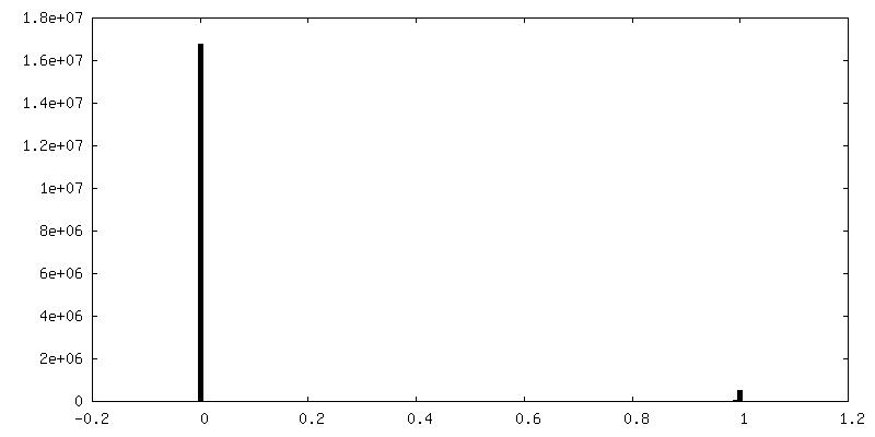
| FSC plot (resolution estimation) |  |

