 Movie
Movie Controller


+ Open data

- Basic information
| Entry | Database: PDB / ID: 8gzw | ||||||
|---|---|---|---|---|---|---|---|
| Title | Klebsiella pneumoniae FtsZ complexed with monobody (P21) | ||||||
 Components Components |
| ||||||
 Keywords Keywords |  CELL CYCLE / Cell division / Monobody / GTPase / HYDROLASE CELL CYCLE / Cell division / Monobody / GTPase / HYDROLASE | ||||||
| Function / homology |  Function and homology information Function and homology informationFtsZ-dependent cytokinesis / division septum assembly / cell division site / protein polymerization / GTPase activity / GTP binding / cytoplasmSimilarity search - Function | ||||||
| Biological species |  Klebsiella pneumoniae (bacteria) Klebsiella pneumoniae (bacteria) Homo sapiens (human) Homo sapiens (human) | ||||||
| Method | X-RAY DIFFRACTION / SYNCHROTRON / MOLECULAR REPLACEMENT / Resolution: 2.5 Å | ||||||
 Authors Authors | Matsumura, H. / Yoshizawa, T. / Fujita, J. / Tanaka, S. / Amesaka, H. | ||||||
| Funding support |  Japan, 1items Japan, 1items
| ||||||
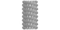
 Citation Citation | Journal: Nat Commun / Year: 2023 Title: Structures of a FtsZ single protofilament and a double-helical tube in complex with a monobody. Authors: Junso Fujita / Hiroshi Amesaka / Takuya Yoshizawa / Kota Hibino / Natsuki Kamimura / Natsuko Kuroda / Takamoto Konishi / Yuki Kato / Mizuho Hara / Tsuyoshi Inoue / Keiichi Namba / Shun-Ichi ...Authors: Junso Fujita / Hiroshi Amesaka / Takuya Yoshizawa / Kota Hibino / Natsuki Kamimura / Natsuko Kuroda / Takamoto Konishi / Yuki Kato / Mizuho Hara / Tsuyoshi Inoue / Keiichi Namba / Shun-Ichi Tanaka / Hiroyoshi Matsumura / Abstract: FtsZ polymerizes into protofilaments to form the Z-ring that acts as a scaffold for accessory proteins during cell division. Structures of FtsZ have been previously solved, but detailed mechanistic ...FtsZ polymerizes into protofilaments to form the Z-ring that acts as a scaffold for accessory proteins during cell division. Structures of FtsZ have been previously solved, but detailed mechanistic insights are lacking. Here, we determine the cryoEM structure of a single protofilament of FtsZ from Klebsiella pneumoniae (KpFtsZ) in a polymerization-preferred conformation. We also develop a monobody (Mb) that binds to KpFtsZ and FtsZ from Escherichia coli without affecting their GTPase activity. Crystal structures of the FtsZ-Mb complexes reveal the Mb binding mode, while addition of Mb in vivo inhibits cell division. A cryoEM structure of a double-helical tube of KpFtsZ-Mb at 2.7 Å resolution shows two parallel protofilaments. Our present study highlights the physiological roles of the conformational changes of FtsZ in treadmilling that regulate cell division. | ||||||
| History |
|
- Structure visualization
| Structure viewer | Molecule: MolmilJmol/JSmol |
|---|
- Downloads & links
-Download
| PDBx/mmCIF format | 8gzw.cif.gz | 277.7 KB | Display | PDBx/mmCIF format |
|---|---|---|---|---|
| PDB format | pdb8gzw.ent.gz | 180.3 KB | Display | PDB format |
| PDBx/mmJSON format | 8gzw.json.gz | Tree view | PDBx/mmJSON format | |
| Others |  Other downloads Other downloads |
-Validation report
| Arichive directory | https://data.pdbj.org/pub/pdb/validation_reports/gz/8gzwftp://data.pdbj.org/pub/pdb/validation_reports/gz/8gzw | HTTPS FTP |
|---|
-Related structure data
| Related structure data | 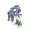 8gzvC  8gzxC  8gzyC 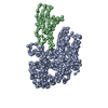 8h1oC 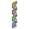 8ibnC  6ll5S S: Starting model for refinement C: citing same article ( |
|---|---|
| Similar structure data |
-Links
 PDBj
PDBj


- Assembly
| Deposited unit | 
| ||||||||||||
|---|---|---|---|---|---|---|---|---|---|---|---|---|---|
| 1 | 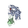
| ||||||||||||
| 2 | 
| ||||||||||||
| 3 | 
| ||||||||||||
| Unit cell |
|
-Components
| #1: Protein | / Filamenting temperature-sensitive mutant Z Mass: 31813.107 Da / Num. of mol.: 3 Source method: isolated from a genetically manipulated source Source: (gene. exp.) Klebsiella pneumoniae (bacteria) / Gene: ftsZ / Production host: Escherichia coli (E. coli) / References: UniProt: W9BCK7#2: Antibody | Mass: 9781.873 Da / Num. of mol.: 3 Source method: isolated from a genetically manipulated source Source: (gene. exp.) Homo sapiens (human) / Production host: Escherichia coli (E. coli)#3: Chemical | Guanosine diphosphate  Type: RNA linking / Mass: 443.201 Da / Num. of mol.: 3 / Source method: obtained synthetically / Formula: C10H15N5O11P2 / Feature type: SUBJECT OF INVESTIGATION / Comment: GDP, energy-carrying molecule*YM Type: RNA linking / Mass: 443.201 Da / Num. of mol.: 3 / Source method: obtained synthetically / Formula: C10H15N5O11P2 / Feature type: SUBJECT OF INVESTIGATION / Comment: GDP, energy-carrying molecule*YM#4: Water | ChemComp-HOH / | Water Mass: 18.015 Da / Num. of mol.: 40 / Source method: isolated from a natural source / Formula: H2O Mass: 18.015 Da / Num. of mol.: 40 / Source method: isolated from a natural source / Formula: H2OHas ligand of interest | Y | |
|---|
-Experimental details
-Experiment
| Experiment | Method: X-RAY DIFFRACTION / Number of used crystals: 1 |
|---|
- Sample preparation
| Crystal | Density Matthews: 2.42 Å3/Da / Density % sol: 49.23 % |
|---|---|
| Crystal grow | Temperature: 293 K / Method: vapor diffusion Details: 0.2M sodium formate, 0.1M Bis-Tris propane pH6.5, 20% PEG 3350 |
-Data collection
| Diffraction | Mean temperature: 100 K / Serial crystal experiment: N |
|---|---|
| Diffraction source | Source: SYNCHROTRON / Site: SPring-8 / Beamline: BL44XU / Wavelength: 0.9 Å |
| Detector | Type: DECTRIS EIGER X 16M / Detector: PIXEL / Date: Jul 24, 2021 |
| Radiation | Protocol: SINGLE WAVELENGTH / Monochromatic (M) / Laue (L): M / Scattering type: x-ray |
| Radiation wavelength | Wavelength: 0.9 Å / Relative weight: 1 |
| Reflection | Resolution: 2.5→40.05 Å / Num. obs: 41534 / % possible obs: 99.69 % / Redundancy: 6.9 % / Biso Wilson estimate: 55.89 Å2 / CC1/2: 0.998 / Net I/σ(I): 15.57 |
| Reflection shell | Resolution: 2.5→2.589 Å / Num. unique obs: 4148 / CC1/2: 0.866 |
- Processing
| Software |
| ||||||||||||||||||||||||||||||||||||||||||||||||||||||||||||||||||||||||||||||||||||||||||||||||||||||||||||||||
|---|---|---|---|---|---|---|---|---|---|---|---|---|---|---|---|---|---|---|---|---|---|---|---|---|---|---|---|---|---|---|---|---|---|---|---|---|---|---|---|---|---|---|---|---|---|---|---|---|---|---|---|---|---|---|---|---|---|---|---|---|---|---|---|---|---|---|---|---|---|---|---|---|---|---|---|---|---|---|---|---|---|---|---|---|---|---|---|---|---|---|---|---|---|---|---|---|---|---|---|---|---|---|---|---|---|---|---|---|---|---|---|---|---|
| Refinement | Method to determine structure: MOLECULAR REPLACEMENT Starting model: 6LL5 Resolution: 2.5→40.05 Å / SU ML: 0.4442 / Cross valid method: FREE R-VALUE / σ(F): 1.36 / Phase error: 32.3394 Stereochemistry target values: GeoStd + Monomer Library + CDL v1.2
| ||||||||||||||||||||||||||||||||||||||||||||||||||||||||||||||||||||||||||||||||||||||||||||||||||||||||||||||||
| Solvent computation | Shrinkage radii: 0.9 Å / VDW probe radii: 1.11 Å / Solvent model: FLAT BULK SOLVENT MODEL | ||||||||||||||||||||||||||||||||||||||||||||||||||||||||||||||||||||||||||||||||||||||||||||||||||||||||||||||||
| Displacement parameters | Biso mean: 57.11 Å2 | ||||||||||||||||||||||||||||||||||||||||||||||||||||||||||||||||||||||||||||||||||||||||||||||||||||||||||||||||
| Refinement step | Cycle: LAST / Resolution: 2.5→40.05 Å
| ||||||||||||||||||||||||||||||||||||||||||||||||||||||||||||||||||||||||||||||||||||||||||||||||||||||||||||||||
| Refine LS restraints |
| ||||||||||||||||||||||||||||||||||||||||||||||||||||||||||||||||||||||||||||||||||||||||||||||||||||||||||||||||
| LS refinement shell |
|
