Movie
Movie Controller


+ Open data

- Basic information
| Entry | Database: PDB / ID: 8b0h | |||||||||
|---|---|---|---|---|---|---|---|---|---|---|
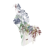
| Title | 2C9, C5b9-CD59 cryoEM structure | |||||||||
 Components Components |
| |||||||||
 Keywords Keywords |  MEMBRANE PROTEIN / Complement / CD59 / MAC MEMBRANE PROTEIN / Complement / CD59 / MAC | |||||||||
| Function / homology |  Function and homology information Function and homology informationnegative regulation of activation of membrane attack complex / cell killing / Terminal pathway of complement / membrane attack complex / complement binding / regulation of complement-dependent cytotoxicity / other organism cell membrane / regulation of complement activation / Activation of C3 and C5 / negative regulation of macrophage chemotaxis ...negative regulation of activation of membrane attack complex / cell killing / Terminal pathway of complement / membrane attack complex / complement binding / regulation of complement-dependent cytotoxicity / other organism cell membrane / regulation of complement activation / Activation of C3 and C5 / negative regulation of macrophage chemotaxis / Cargo concentration in the ER / complement activation, alternative pathway / complement activation / chemokine activity / COPII-mediated vesicle transport / retinol binding / endopeptidase inhibitor activity / tertiary granule membrane / positive regulation of vascular endothelial growth factor production / specific granule membrane / COPI-mediated anterograde transport / complement activation, classical pathway / positive regulation of chemokine production / transport vesicle / endoplasmic reticulum-Golgi intermediate compartment membrane / Peptide ligand-binding receptors / Regulation of Complement cascade / protein homooligomerization / ER to Golgi transport vesicle membrane / chemotaxis / positive regulation of immune response / extracellular vesicle / blood coagulation / G alpha (i) signalling events / blood microparticle / killing of cells of another organism / in utero embryonic development / vesicle / cell surface receptor signaling pathway / immune response / inflammatory response / G protein-coupled receptor signaling pathway / external side of plasma membrane / Golgi membrane / signaling receptor binding / focal adhesion / innate immune response / Neutrophil degranulation / protein-containing complex binding / endoplasmic reticulum membrane / cell surface / extracellular space / extracellular exosome / extracellular region / membrane / plasma membraneSimilarity search - Function | |||||||||
| Biological species |  Homo sapiens (human) Homo sapiens (human) | |||||||||
| Method | ELECTRON MICROSCOPY / single particle reconstruction / cryo EM / Resolution: 3.3 Å | |||||||||
 Authors Authors | Couves, E.C. / Gardner, S. / Bubeck, D. | |||||||||
| Funding support | European Union,  United Kingdom, 2items United Kingdom, 2items
| |||||||||
 Citation Citation | Journal: Nat Commun / Year: 2023 Title: Structural basis for membrane attack complex inhibition by CD59. Authors: Emma C Couves / Scott Gardner / Tomas B Voisin / Jasmine K Bickel / Phillip J Stansfeld / Edward W Tate / Doryen Bubeck / Abstract: CD59 is an abundant immuno-regulatory receptor that protects human cells from damage during complement activation. Here we show how the receptor binds complement proteins C8 and C9 at the membrane to ...CD59 is an abundant immuno-regulatory receptor that protects human cells from damage during complement activation. Here we show how the receptor binds complement proteins C8 and C9 at the membrane to prevent insertion and polymerization of membrane attack complex (MAC) pores. We present cryo-electron microscopy structures of two inhibited MAC precursors known as C5b8 and C5b9. We discover that in both complexes, CD59 binds the pore-forming β-hairpins of C8 to form an intermolecular β-sheet that prevents membrane perforation. While bound to C8, CD59 deflects the cascading C9 β-hairpins, rerouting their trajectory into the membrane. Preventing insertion of C9 restricts structural transitions of subsequent monomers and indirectly halts MAC polymerization. We combine our structural data with cellular assays and molecular dynamics simulations to explain how the membrane environment impacts the dual roles of CD59 in controlling pore formation of MAC, and as a target of bacterial virulence factors which hijack CD59 to lyse human cells. | |||||||||
| History |
|
- Structure visualization
| Structure viewer | Molecule: MolmilJmol/JSmol |
|---|
- Downloads & links
-Download
| PDBx/mmCIF format | 8b0h.cif.gz | 1.5 MB | Display | PDBx/mmCIF format |
|---|---|---|---|---|
| PDB format | pdb8b0h.ent.gz | Display | PDB format | |
| PDBx/mmJSON format | 8b0h.json.gz | Tree view | PDBx/mmJSON format | |
| Others |  Other downloads Other downloads |
-Validation report
| Arichive directory | https://data.pdbj.org/pub/pdb/validation_reports/b0/8b0hftp://data.pdbj.org/pub/pdb/validation_reports/b0/8b0h | HTTPS FTP |
|---|
-Related structure data
| Related structure data |  15781MC 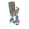 8b0fC 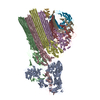 8b0gC M: map data used to model this data C: citing same article ( |
|---|---|
| Similar structure data |


-Links
 PDBj
PDBj


- Assembly
| Deposited unit | 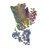
|
|---|---|
| 1 |
|
-Components
-Protein , 2 types, 2 molecules GA
| #1: Protein | Mass: 14234.388 Da / Num. of mol.: 1 Source method: isolated from a genetically manipulated source Source: (gene. exp.) Homo sapiens (human) / Gene: CD59, MIC11, MIN1, MIN2, MIN3, MSK21 / Production host:  Escherichia coli BL21 (bacteria) / References: UniProt: P13987 Escherichia coli BL21 (bacteria) / References: UniProt: P13987 |
|---|---|
| #5: Protein | Complement component 5 / C3 and PZP-like alpha-2-macroglobulin domain-containing protein 4 Mass: 188512.094 Da / Num. of mol.: 1 / Source method: isolated from a natural source / Source: (natural) Homo sapiens (human) / References: UniProt: P01031 |
-Complement component ... , 6 types, 7 molecules DFECBHI
| #2: Protein | Mass: 67136.891 Da / Num. of mol.: 1 / Source method: isolated from a natural source / Source: (natural) Homo sapiens (human) / References: UniProt: P07358 |
|---|---|
| #3: Protein | Mass: 22302.424 Da / Num. of mol.: 1 / Source method: isolated from a natural source / Source: (natural) Homo sapiens (human) / References: UniProt: P07360 |
| #4: Protein | Mass: 65239.152 Da / Num. of mol.: 1 / Source method: isolated from a natural source / Source: (natural) Homo sapiens (human) / References: UniProt: P07357 |
| #6: Protein | Mass: 93625.328 Da / Num. of mol.: 1 / Source method: isolated from a natural source / Source: (natural) Homo sapiens (human) / References: UniProt: P10643 |
| #7: Protein | Mass: 104918.180 Da / Num. of mol.: 1 / Source method: isolated from a natural source / Source: (natural) Homo sapiens (human) / References: UniProt: P13671 |
| #8: Protein | Mass: 63252.301 Da / Num. of mol.: 2 / Source method: isolated from a natural source / Source: (natural) Homo sapiens (human) / References: UniProt: P02748 |
-Experimental details
-Experiment
| Experiment | Method: ELECTRON MICROSCOPY |
|---|---|
| EM experiment | Aggregation state: PARTICLE / 3D reconstruction method: single particle reconstruction |
- Sample preparation
| Component |
| ||||||||||||||||||||||||||||
|---|---|---|---|---|---|---|---|---|---|---|---|---|---|---|---|---|---|---|---|---|---|---|---|---|---|---|---|---|---|
| Molecular weight | Experimental value: NO | ||||||||||||||||||||||||||||
| Source (natural) |
| ||||||||||||||||||||||||||||
| Source (recombinant) | Organism: Escherichia coli BL21 (bacteria) | ||||||||||||||||||||||||||||
| Buffer solution | pH: 7.4 / Details: 20 mM HEPES pH 7.4, 120 mM NaCl | ||||||||||||||||||||||||||||
| Buffer component |
| ||||||||||||||||||||||||||||
| Specimen | Embedding applied: NO / Shadowing applied: NO / Staining applied: NO / Vitrification applied: YES | ||||||||||||||||||||||||||||
| Specimen support | Grid material: COPPER / Grid mesh size: 300 divisions/in. / Grid type: Quantifoil R1.2/1.3 | ||||||||||||||||||||||||||||
| Vitrification | Instrument: FEI VITROBOT MARK II / Cryogen name: ETHANE / Humidity: 95 % |
- Electron microscopy imaging
| Experimental equipment |  Model: Titan Krios / Image courtesy: FEI Company |
|---|---|
| Microscopy | Model: FEI TITAN KRIOS |
| Electron gun | Electron source: FIELD EMISSION GUN / Accelerating voltage: 300 kV / Illumination mode: FLOOD BEAM |
| Electron lens | Mode: BRIGHT FIELDBright-field microscopy / Nominal magnification: 105000 X / Nominal defocus max: 9000 nm / Nominal defocus min: 1000 nm / Calibrated defocus max: 4000 nm / Cs: 2.7 mm / C2 aperture diameter: 100 µm / Alignment procedure: COMA FREE |
| Specimen holder | Specimen holder model: FEI TITAN KRIOS AUTOGRID HOLDER |
| Image recording | Average exposure time: 2 sec. / Electron dose: 50 e/Å2 / Film or detector model: GATAN K3 (6k x 4k) / Num. of grids imaged: 4 / Num. of real images: 52838 / Details: Collected over 4 separate data collections |
- Processing
| Software | Name: PHENIX / Version: 1.20_4459: / Classification: refinement | ||||||||||||||||||||||||||||||||||||
|---|---|---|---|---|---|---|---|---|---|---|---|---|---|---|---|---|---|---|---|---|---|---|---|---|---|---|---|---|---|---|---|---|---|---|---|---|---|
| EM software |
| ||||||||||||||||||||||||||||||||||||
| CTF correction | Type: PHASE FLIPPING AND AMPLITUDE CORRECTION | ||||||||||||||||||||||||||||||||||||
| Particle selection | Details: Dataset 1: 737,138 Dataset 2: 1,058,026 Dataset 3: 1,330,232 Dataset 4: 722,870 | ||||||||||||||||||||||||||||||||||||
| 3D reconstruction | Resolution: 3.3 Å / Resolution method: FSC 0.143 CUT-OFF / Num. of particles: 47244 / Symmetry type: POINT | ||||||||||||||||||||||||||||||||||||
| Atomic model building | Details: Initial fitting was done in ISODLE, final refinements were done in ISOLDE | ||||||||||||||||||||||||||||||||||||
| Atomic model building |
| ||||||||||||||||||||||||||||||||||||
| Refine LS restraints |
|