ムービー
ムービー コントローラー


+ データを開く

- 基本情報
基本情報
| 登録情報 | 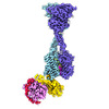 | |||||||||
|---|---|---|---|---|---|---|---|---|---|---|
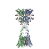
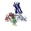
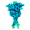
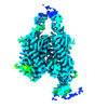
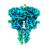
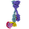
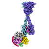
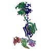
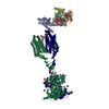
| タイトル | Structure of human calcium-sensing receptor in complex with chimeric Gs (miniGis) protein in nanodiscs | |||||||||
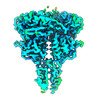
 マップデータ マップデータ | Structure of human calcium-sensing receptor in complex with chimeric Gs (miniGis) protein | |||||||||
 試料 試料 |
| |||||||||
 キーワード キーワード |  Calcium-sensing receptor (カルシウム感知受容体) / G-protein-coupled receptor (Gタンパク質共役受容体) / G protein (Gタンパク質) / signal transduction (シグナル伝達) / MEMBRANE PROTEIN (膜タンパク質) Calcium-sensing receptor (カルシウム感知受容体) / G-protein-coupled receptor (Gタンパク質共役受容体) / G protein (Gタンパク質) / signal transduction (シグナル伝達) / MEMBRANE PROTEIN (膜タンパク質) | |||||||||
| 機能・相同性 |  機能・相同性情報 機能・相同性情報bile acid secretion / chemosensory behavior / cellular response to peptide / response to fibroblast growth factor / cellular response to vitamin D / phosphatidylinositol phospholipase C activity / Class C/3 (Metabotropic glutamate/pheromone receptors) / calcium ion import / positive regulation of positive chemotaxis / fat pad development ...bile acid secretion / chemosensory behavior / cellular response to peptide / response to fibroblast growth factor / cellular response to vitamin D / phosphatidylinositol phospholipase C activity / Class C/3 (Metabotropic glutamate/pheromone receptors) / calcium ion import / positive regulation of positive chemotaxis / fat pad development / amino acid binding / cellular response to hepatocyte growth factor stimulus / branching morphogenesis of an epithelial tube / positive regulation of calcium ion import / regulation of calcium ion transport / cellular response to low-density lipoprotein particle stimulus / detection of calcium ion / Adenylate cyclase inhibitory pathway / positive regulation of protein localization to cell cortex / anatomical structure morphogenesis / regulation of cAMP-mediated signaling / D2 dopamine receptor binding / G protein-coupled serotonin receptor binding / axon terminus / regulation of mitotic spindle organization / cellular response to forskolin / JNK cascade / positive regulation of vasoconstriction / chloride transmembrane transport / adenylate cyclase-inhibiting G protein-coupled receptor signaling pathway / 骨化 / Regulation of insulin secretion / G protein-coupled receptor binding / response to ischemia / G protein-coupled receptor activity / cellular response to glucose stimulus / Olfactory Signaling Pathway / G-protein beta/gamma-subunit complex binding / Activation of the phototransduction cascade / G beta:gamma signalling through PLC beta / Presynaptic function of Kainate receptors / Thromboxane signalling through TP receptor / adenylate cyclase-modulating G protein-coupled receptor signaling pathway / G-protein activation / G protein-coupled acetylcholine receptor signaling pathway / Activation of G protein gated Potassium channels / Inhibition of voltage gated Ca2+ channels via Gbeta/gamma subunits / Prostacyclin signalling through prostacyclin receptor / Glucagon signaling in metabolic regulation / G beta:gamma signalling through CDC42 / positive regulation of insulin secretion / ADP signalling through P2Y purinoceptor 12 / G beta:gamma signalling through BTK / Synthesis, secretion, and inactivation of Glucagon-like Peptide-1 (GLP-1) / Sensory perception of sweet, bitter, and umami (glutamate) taste / photoreceptor disc membrane / response to peptide hormone / Adrenaline,noradrenaline inhibits insulin secretion / Glucagon-type ligand receptors / Vasopressin regulates renal water homeostasis via Aquaporins / intracellular calcium ion homeostasis / G alpha (z) signalling events / cellular response to catecholamine stimulus / Glucagon-like Peptide-1 (GLP1) regulates insulin secretion / ADORA2B mediated anti-inflammatory cytokines production / adenylate cyclase-activating dopamine receptor signaling pathway / ADP signalling through P2Y purinoceptor 1 / G beta:gamma signalling through PI3Kgamma / cellular response to prostaglandin E stimulus / Cooperation of PDCL (PhLP1) and TRiC/CCT in G-protein beta folding / vasodilation / sensory perception of taste / GPER1 signaling / G-protein beta-subunit binding / GDP binding / Inactivation, recovery and regulation of the phototransduction cascade / heterotrimeric G-protein complex / G alpha (12/13) signalling events / extracellular vesicle / signaling receptor complex adaptor activity / Thrombin signalling through proteinase activated receptors (PARs) / integrin binding / retina development in camera-type eye / GTPase binding / Ca2+ pathway / phospholipase C-activating G protein-coupled receptor signaling pathway / 細胞皮質 / midbody / G alpha (i) signalling events / fibroblast proliferation / cellular response to hypoxia / G alpha (s) signalling events / G alpha (q) signalling events / basolateral plasma membrane / Ras protein signal transduction / cell population proliferation / transmembrane transporter binding / Extra-nuclear estrogen signaling / positive regulation of ERK1 and ERK2 cascade / 細胞周期類似検索 - 分子機能 | |||||||||
| 生物種 |  Homo sapiens (ヒト) Homo sapiens (ヒト) | |||||||||
| 手法 | 単粒子再構成法 / クライオ電子顕微鏡法 / 解像度: 3.6 Å | |||||||||
 データ登録者 データ登録者 | Zuo H / Park J / Frangaj A / Ye J / Lu G / Manning JJ / Asher WB / Lu Z / Hu G / Wang L ...Zuo H / Park J / Frangaj A / Ye J / Lu G / Manning JJ / Asher WB / Lu Z / Hu G / Wang L / Mendez J / Eng E / Zhang Z / Lin X / Grasucci R / Hendrickson WA / Clarke OB / Javitch JA / Conigrave AD / Fan QR | |||||||||
| 資金援助 |  米国, 1件 米国, 1件
| |||||||||
 引用 引用 | ジャーナル: Nature / 年: 2024 タイトル: Promiscuous G-protein activation by the calcium-sensing receptor. 著者: Hao Zuo / Jinseo Park / Aurel Frangaj / Jianxiang Ye / Guanqi Lu / Jamie J Manning / Wesley B Asher / Zhengyuan Lu / Guo-Bin Hu / Liguo Wang / Joshua Mendez / Edward Eng / Zhening Zhang / Xin ...著者: Hao Zuo / Jinseo Park / Aurel Frangaj / Jianxiang Ye / Guanqi Lu / Jamie J Manning / Wesley B Asher / Zhengyuan Lu / Guo-Bin Hu / Liguo Wang / Joshua Mendez / Edward Eng / Zhening Zhang / Xin Lin / Robert Grassucci / Wayne A Hendrickson / Oliver B Clarke / Jonathan A Javitch / Arthur D Conigrave / Qing R Fan /  要旨: The human calcium-sensing receptor (CaSR) detects fluctuations in the extracellular Ca concentration and maintains Ca homeostasis. It also mediates diverse cellular processes not associated with Ca ...The human calcium-sensing receptor (CaSR) detects fluctuations in the extracellular Ca concentration and maintains Ca homeostasis. It also mediates diverse cellular processes not associated with Ca balance. The functional pleiotropy of CaSR arises in part from its ability to signal through several G-protein subtypes. We determined structures of CaSR in complex with G proteins from three different subfamilies: G, G and G. We found that the homodimeric CaSR of each complex couples to a single G protein through a common mode. This involves the C-terminal helix of each Gα subunit binding to a shallow pocket that is formed in one CaSR subunit by all three intracellular loops (ICL1-ICL3), an extended transmembrane helix 3 and an ordered C-terminal region. G-protein binding expands the transmembrane dimer interface, which is further stabilized by phospholipid. The restraint imposed by the receptor dimer, in combination with ICL2, enables G-protein activation by facilitating conformational transition of Gα. We identified a single Gα residue that determines G and G versus G selectivity. The length and flexibility of ICL2 allows CaSR to bind all three Gα subtypes, thereby conferring capacity for promiscuous G-protein coupling. | |||||||||
| 履歴 |
|
- 構造の表示
構造の表示
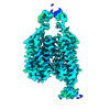
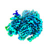
| 添付画像 |
|---|

- ダウンロードとリンク
-EMDBアーカイブ
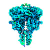
| マップデータ | emd_43901.map.gz | 10.9 MB | EMDBマップデータ形式 | |
|---|---|---|---|---|
| ヘッダ (付随情報) | emd-43901-v30.xmlemd-43901.xml | 27.4 KB 27.4 KB | 表示 表示 | EMDBヘッダ |
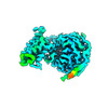
| 画像 |  emd_43901.png emd_43901.png | 100.1 KB | ||
| Filedesc metadata | emd-43901.cif.gz | 8.5 KB | ||
| アーカイブディレクトリ |  http://ftp.pdbj.org/pub/emdb/structures/EMD-43901ftp://ftp.pdbj.org/pub/emdb/structures/EMD-43901 http://ftp.pdbj.org/pub/emdb/structures/EMD-43901ftp://ftp.pdbj.org/pub/emdb/structures/EMD-43901 | HTTPS FTP |
-関連構造データ


-リンク
| EMDBのページ | EMDB (EBI/PDBe) / EMDataResource |
|---|---|
| 「今月の分子」の関連する項目 |


-マップ
| ファイル | ダウンロード / ファイル: emd_43901.map.gz / 形式: CCP4 / 大きさ: 512 MB / タイプ: IMAGE STORED AS FLOATING POINT NUMBER (4 BYTES) | ||||||||||||||||||||
|---|---|---|---|---|---|---|---|---|---|---|---|---|---|---|---|---|---|---|---|---|---|
| 注釈 | Structure of human calcium-sensing receptor in complex with chimeric Gs (miniGis) protein | ||||||||||||||||||||
| ボクセルのサイズ | X=Y=Z: 0.825 Å | ||||||||||||||||||||
| 密度 |
| ||||||||||||||||||||
| 対称性 | 空間群: 1 | ||||||||||||||||||||
| 詳細 | EMDB XML:
|
-添付データ
- 試料の構成要素
+全体 : Human CaSR in complex with chimeric Gs (miniGis) protein
+超分子 #1: Human CaSR in complex with chimeric Gs (miniGis) protein
+分子 #1: Isoform 1 of Extracellular calcium-sensing receptor
+分子 #2: Chimeric mini guanine nucleotide-binding protein G(i)(s) subunit alpha
+分子 #3: Guanine nucleotide-binding protein G(I)/G(S)/G(T) subunit beta-1
+分子 #4: Guanine nucleotide-binding protein G(I)/G(S)/G(O) subunit gamma-2
+分子 #6: 2-acetamido-2-deoxy-beta-D-glucopyranose

+分子 #7: CYCLOMETHYLTRYPTOPHAN
+分子 #8: PHOSPHATE ION

+分子 #9: CALCIUM ION
+分子 #10: 3-(2-chlorophenyl)-N-[(1R)-1-(3-methoxyphenyl)ethyl]propan-1-amine

+分子 #11: CHOLESTEROL HEMISUCCINATE

+分子 #12: (19R,22S,25R)-22,25,26-trihydroxy-16,22-dioxo-17,21,23-trioxa-22l...
-実験情報
-構造解析
| 手法 | クライオ電子顕微鏡法 |
|---|---|
 解析 解析 | 単粒子再構成法 |
| 試料の集合状態 | particle |
-試料調製
| 濃度 | 3.9 mg/mL | |||||||||||||||||||||
|---|---|---|---|---|---|---|---|---|---|---|---|---|---|---|---|---|---|---|---|---|---|---|
| 緩衝液 | pH: 7.5 構成要素:
| |||||||||||||||||||||
| グリッド | モデル: Quantifoil R0.6/1 / 材質: GOLD / メッシュ: 300 / 支持フィルム - 材質: GOLD / 支持フィルム - トポロジー: HOLEY / 支持フィルム - Film thickness: 50 / 前処理 - タイプ: GLOW DISCHARGE / 前処理 - 時間: 25 sec. / 前処理 - 雰囲気: OTHER | |||||||||||||||||||||
| 凍結 | 凍結剤: ETHANE / チャンバー内湿度: 100 % / チャンバー内温度: 277 K / 装置: FEI VITROBOT MARK IV 詳細: The sample was blotted for 6s before plunge-frozen.. |
- 電子顕微鏡法
| 顕微鏡 | FEI TITAN KRIOS |
|---|---|
| 電子線 | 加速電圧: 300 kV / 電子線源: FIELD EMISSION GUN |
| 電子光学系 | C2レンズ絞り径: 100.0 µm / 照射モード: FLOOD BEAM / 撮影モード: BRIGHT FIELDBright-field microscopy / Cs: 2.7 mm 最大 デフォーカス(公称値): 1.9000000000000001 µm 最小 デフォーカス(公称値): 1.1 µm / 倍率(公称値): 105000 |
| 特殊光学系 | エネルギーフィルター - 名称: GIF Bioquantum / エネルギーフィルター - スリット幅: 15 eV |
| 試料ステージ | 試料ホルダーモデル: FEI TITAN KRIOS AUTOGRID HOLDER ホルダー冷却材: NITROGEN |
| 温度 | 最高: 100.0 K |
| ソフトウェア | 名称: EPU |
| 撮影 | フィルム・検出器のモデル: GATAN K3 BIOQUANTUM (6k x 4k) デジタル化 - サイズ - 横: 11520 pixel / デジタル化 - サイズ - 縦: 8184 pixel / 撮影したグリッド数: 1 / 実像数: 29091 / 平均露光時間: 2.72 sec. / 平均電子線量: 67.9 e/Å2 |
| 実験機器 |  モデル: Titan Krios / 画像提供: FEI Company |
-画像解析
| 粒子像選択 | 選択した数: 4867087 |
|---|---|
| 初期モデル | モデルのタイプ: OTHER / 詳細: Ab initio |
| 初期 角度割当 | タイプ: MAXIMUM LIKELIHOOD / ソフトウェア - 名称: cryoSPARC (ver. 3.3.2) |
| 最終 角度割当 | タイプ: MAXIMUM LIKELIHOOD / ソフトウェア - 名称: cryoSPARC (ver. 3.3.2) |
| 最終 再構成 | 想定した対称性 - 点群: C1 (非対称) / 解像度のタイプ: BY AUTHOR / 解像度: 3.6 Å / 解像度の算出法: FSC 0.143 CUT-OFF / ソフトウェア - 名称: cryoSPARC (ver. 3.3.2) / 使用した粒子像数: 45862 |
-原子モデル構築 1
| 初期モデル |
| ||||||||||||
|---|---|---|---|---|---|---|---|---|---|---|---|---|---|
| ソフトウェア | 名称: Coot (ver. 0.9.8.1) | ||||||||||||
| 精密化 | 空間: REAL / プロトコル: FLEXIBLE FIT | ||||||||||||
| 得られたモデル |  PDB-9avg: |