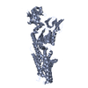
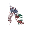
Movie
Movie Controller


+ Open data

- Basic information
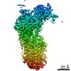
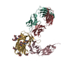
| Entry | Database: PDB / ID: 7si3 | |||||||||
|---|---|---|---|---|---|---|---|---|---|---|
| Title | Consensus structure of ATP7B | |||||||||
 Components Components | P-type Cu(+) transporter | |||||||||
 Keywords Keywords | METAL TRANSPORT/Translocase / Copper transport /  Wilson disease / METAL TRANSPORT / METAL TRANSPORT-Translocase complex Wilson disease / METAL TRANSPORT / METAL TRANSPORT-Translocase complex | |||||||||
| Function / homology |  Function and homology information Function and homology informationIon transport by P-type ATPases / P-type divalent copper transporter activity / P-type monovalent copper transporter activity / P-type Cu+ transporter / copper ion export / copper ion import / intracellular copper ion homeostasis / trans-Golgi network / copper ion binding / ATP hydrolysis activity ...Ion transport by P-type ATPases / P-type divalent copper transporter activity / P-type monovalent copper transporter activity / P-type Cu+ transporter / copper ion export / copper ion import / intracellular copper ion homeostasis / trans-Golgi network / copper ion binding / ATP hydrolysis activity / ATP binding / plasma membraneSimilarity search - Function | |||||||||
| Biological species | Xenopus tropicalis (tropical clawed frog) | |||||||||
| Method | ELECTRON MICROSCOPY / single particle reconstruction / cryo EM / Resolution: 3.19 Å | |||||||||
 Authors Authors | Bitter, R.M. / Oh, S.C. / Hite, R.K. / Yuan, P. | |||||||||
| Funding support |  United States, 2items United States, 2items
| |||||||||
 Citation Citation | Journal: Sci Adv / Year: 2022 Title: Structure of the Wilson disease copper transporter ATP7B. Authors: Ryan M Bitter / SeCheol Oh / Zengqin Deng / Suhaila Rahman / Richard K Hite / Peng Yuan / Abstract: ATP7A and ATP7B, two homologous copper-transporting P1B-type ATPases, play crucial roles in cellular copper homeostasis, and mutations cause Menkes and Wilson diseases, respectively. ATP7A/B contains ...ATP7A and ATP7B, two homologous copper-transporting P1B-type ATPases, play crucial roles in cellular copper homeostasis, and mutations cause Menkes and Wilson diseases, respectively. ATP7A/B contains a P-type ATPase core consisting of a membrane transport domain and three cytoplasmic domains, the A, P, and N domains, and a unique amino terminus comprising six consecutive metal-binding domains. Here, we present a cryo-electron microscopy structure of frog ATP7B in a copper-free state. Interacting with both the A and P domains, the metal-binding domains are poised to exert copper-dependent regulation of ATP hydrolysis coupled to transmembrane copper transport. A ring of negatively charged residues lines the cytoplasmic copper entrance that is presumably gated by a conserved basic residue sitting at the center. Within the membrane, a network of copper-coordinating ligands delineates a stepwise copper transport pathway. This work provides the first glimpse into the structure and function of ATP7 proteins and facilitates understanding of disease mechanisms and development of rational therapies. | |||||||||
| History |
|
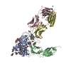
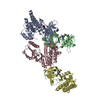
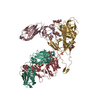
- Structure visualization
| Movie |
Movie viewer |
|---|---|
| Structure viewer | Molecule: MolmilJmol/JSmol |
- Downloads & links
-Download
| PDBx/mmCIF format | 7si3.cif.gz | 297.6 KB | Display | PDBx/mmCIF format |
|---|---|---|---|---|
| PDB format | pdb7si3.ent.gz | 244 KB | Display | PDB format |
| PDBx/mmJSON format | 7si3.json.gz | Tree view | PDBx/mmJSON format | |
| Others |  Other downloads Other downloads |
-Validation report
| Arichive directory | https://data.pdbj.org/pub/pdb/validation_reports/si/7si3ftp://data.pdbj.org/pub/pdb/validation_reports/si/7si3 | HTTPS FTP |
|---|
-Related structure data
| Related structure data | 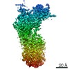 25137MC  7si6C  7si7C M: map data used to model this data C: citing same article ( |
|---|---|
| Similar structure data |


-Links
 PDBj
PDBj


- Assembly
| Deposited unit | 
|
|---|---|
| 1 |
|
-Components
| #1: Protein | Mass: 159678.672 Da / Num. of mol.: 1 Source method: isolated from a genetically manipulated source Source: (gene. exp.) Xenopus tropicalis (tropical clawed frog)Gene: atp7b / Production host:  Komagataella pastoris (fungus) / References: UniProt: A0A6I8R0A5, P-type Cu+ transporter Komagataella pastoris (fungus) / References: UniProt: A0A6I8R0A5, P-type Cu+ transporter |
|---|---|
| #2: Chemical | ChemComp-MG /   Mass: 24.305 Da / Num. of mol.: 1 / Source method: obtained synthetically / Formula: Mg Mass: 24.305 Da / Num. of mol.: 1 / Source method: obtained synthetically / Formula: Mg |
| #3: Chemical | ChemComp-ALF /   Mass: 102.975 Da / Num. of mol.: 1 / Source method: obtained synthetically / Formula: AlF4 / Feature type: SUBJECT OF INVESTIGATION Mass: 102.975 Da / Num. of mol.: 1 / Source method: obtained synthetically / Formula: AlF4 / Feature type: SUBJECT OF INVESTIGATION |
| Has ligand of interest | Y |
-Experimental details
-Experiment
| Experiment | Method: ELECTRON MICROSCOPY |
|---|---|
| EM experiment | Aggregation state: PARTICLE / 3D reconstruction method: single particle reconstruction |
- Sample preparation
| Component | Name: ATP7BWilson disease protein / Type: COMPLEX / Entity ID: #1 / Source: RECOMBINANT | ||||||||||||||||||||||||||||||||||||||||
|---|---|---|---|---|---|---|---|---|---|---|---|---|---|---|---|---|---|---|---|---|---|---|---|---|---|---|---|---|---|---|---|---|---|---|---|---|---|---|---|---|---|
| Molecular weight | Experimental value: NO | ||||||||||||||||||||||||||||||||||||||||
| Source (natural) | Organism: Xenopus tropicalis (tropical clawed frog) | ||||||||||||||||||||||||||||||||||||||||
| Source (recombinant) | Organism: Komagataella pastoris (fungus) | ||||||||||||||||||||||||||||||||||||||||
| Buffer solution | pH: 7 | ||||||||||||||||||||||||||||||||||||||||
| Buffer component |
| ||||||||||||||||||||||||||||||||||||||||
| Specimen | Conc.: 7 mg/ml / Embedding applied: NO / Shadowing applied: NO / Staining applied: NO / Vitrification applied: YES | ||||||||||||||||||||||||||||||||||||||||
| Specimen support | Grid material: GOLD / Grid mesh size: 400 divisions/in. / Grid type: Quantifoil R1.2/1.3 | ||||||||||||||||||||||||||||||||||||||||
| Vitrification | Instrument: FEI VITROBOT MARK IV / Cryogen name: ETHANE / Humidity: 100 % / Chamber temperature: 298 K |
- Electron microscopy imaging
| Experimental equipment |  Model: Titan Krios / Image courtesy: FEI Company |
|---|---|
| Microscopy | Model: FEI TITAN KRIOS |
| Electron gun | Electron source: FIELD EMISSION GUN / Accelerating voltage: 300 kV / Illumination mode: FLOOD BEAM |
| Electron lens | Mode: BRIGHT FIELDBright-field microscopy / Alignment procedure: COMA FREE |
| Specimen holder | Cryogen: NITROGEN / Specimen holder model: FEI TITAN KRIOS AUTOGRID HOLDER |
| Image recording | Electron dose: 61 e/Å2 / Detector mode: SUPER-RESOLUTION / Film or detector model: GATAN K2 QUANTUM (4k x 4k) |
- Processing
| Software | Name: PHENIX / Version: 1.19.2_4158: / Classification: refinement | ||||||||||||||||||||||||||||||||||||
|---|---|---|---|---|---|---|---|---|---|---|---|---|---|---|---|---|---|---|---|---|---|---|---|---|---|---|---|---|---|---|---|---|---|---|---|---|---|
| EM software |
| ||||||||||||||||||||||||||||||||||||
| CTF correction | Type: PHASE FLIPPING AND AMPLITUDE CORRECTION | ||||||||||||||||||||||||||||||||||||
| Particle selection | Num. of particles selected: 1889296 | ||||||||||||||||||||||||||||||||||||
| Symmetry | Point symmetry: C1 (asymmetric) | ||||||||||||||||||||||||||||||||||||
| 3D reconstruction | Resolution: 3.19 Å / Resolution method: FSC 0.143 CUT-OFF / Num. of particles: 257208 / Algorithm: FOURIER SPACE / Num. of class averages: 1 / Symmetry type: POINT | ||||||||||||||||||||||||||||||||||||
| Atomic model building | Protocol: AB INITIO MODEL / Space: REAL | ||||||||||||||||||||||||||||||||||||
| Atomic model building | PDB-ID: 3RFU | ||||||||||||||||||||||||||||||||||||
| Refine LS restraints |
|