Movie
Movie Controller


+ Open data

- Basic information
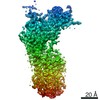
| Entry | Database: EMDB / ID: EMD-25138 | |||||||||
|---|---|---|---|---|---|---|---|---|---|---|
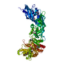
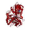
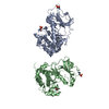
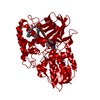
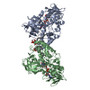
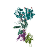
| Title | Structure of ATP7B in state 1 | |||||||||
 Map data Map data | ATP7B State 1 | |||||||||
 Sample Sample |
| |||||||||
| Function / homology |  Function and homology information Function and homology informationP-type monovalent copper transporter activity / P-type Cu+ transporter /  trans-Golgi network / copper ion binding / ATP hydrolysis activity / ATP binding / membrane trans-Golgi network / copper ion binding / ATP hydrolysis activity / ATP binding / membraneSimilarity search - Function | |||||||||
| Biological species | Xenopus tropicalis (tropical clawed frog) | |||||||||
| Method | single particle reconstruction / cryo EM / Resolution: 3.32 Å | |||||||||
 Authors Authors | Bitter RM / Oh SC / Hite RK / Yuan P | |||||||||
| Funding support |  United States, 2 items United States, 2 items
| |||||||||
 Citation Citation | Journal: Sci Adv / Year: 2022 Title: Structure of the Wilson disease copper transporter ATP7B. Authors: Ryan M Bitter / SeCheol Oh / Zengqin Deng / Suhaila Rahman / Richard K Hite / Peng Yuan / Abstract: ATP7A and ATP7B, two homologous copper-transporting P1B-type ATPases, play crucial roles in cellular copper homeostasis, and mutations cause Menkes and Wilson diseases, respectively. ATP7A/B contains ...ATP7A and ATP7B, two homologous copper-transporting P1B-type ATPases, play crucial roles in cellular copper homeostasis, and mutations cause Menkes and Wilson diseases, respectively. ATP7A/B contains a P-type ATPase core consisting of a membrane transport domain and three cytoplasmic domains, the A, P, and N domains, and a unique amino terminus comprising six consecutive metal-binding domains. Here, we present a cryo-electron microscopy structure of frog ATP7B in a copper-free state. Interacting with both the A and P domains, the metal-binding domains are poised to exert copper-dependent regulation of ATP hydrolysis coupled to transmembrane copper transport. A ring of negatively charged residues lines the cytoplasmic copper entrance that is presumably gated by a conserved basic residue sitting at the center. Within the membrane, a network of copper-coordinating ligands delineates a stepwise copper transport pathway. This work provides the first glimpse into the structure and function of ATP7 proteins and facilitates understanding of disease mechanisms and development of rational therapies. | |||||||||
| History |
|
- Structure visualization
| Movie |
Movie viewer |
|---|---|
| Structure viewer | EM map: SurfViewMolmilJmol/JSmol |
| Supplemental images |

- Downloads & links
-EMDB archive
| Map data | emd_25138.map.gz | 8.8 MB | EMDB map data format | |
|---|---|---|---|---|
| Header (meta data) | emd-25138-v30.xmlemd-25138.xml | 15.7 KB 15.7 KB | Display Display | EMDB header |
| Images | 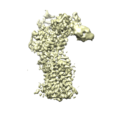 emd_25138.png emd_25138.png | 89.9 KB | ||
| Archive directory |  http://ftp.pdbj.org/pub/emdb/structures/EMD-25138ftp://ftp.pdbj.org/pub/emdb/structures/EMD-25138 http://ftp.pdbj.org/pub/emdb/structures/EMD-25138ftp://ftp.pdbj.org/pub/emdb/structures/EMD-25138 | HTTPS FTP |
-Related structure data
| Related structure data |  7si6MC  7si3C  7si7C M: atomic model generated by this map C: citing same article ( |
|---|---|
| Similar structure data |


-Links
| EMDB pages | EMDB (EBI/PDBe) / EMDataResource |
|---|---|
| Related items in Molecule of the Month |


-Map
| File | Download / File: emd_25138.map.gz / Format: CCP4 / Size: 9.5 MB / Type: IMAGE STORED AS FLOATING POINT NUMBER (4 BYTES) | ||||||||||||||||||||||||||||||||||||||||||||||||||||||||||||||||||||
|---|---|---|---|---|---|---|---|---|---|---|---|---|---|---|---|---|---|---|---|---|---|---|---|---|---|---|---|---|---|---|---|---|---|---|---|---|---|---|---|---|---|---|---|---|---|---|---|---|---|---|---|---|---|---|---|---|---|---|---|---|---|---|---|---|---|---|---|---|---|
| Annotation | ATP7B State 1 | ||||||||||||||||||||||||||||||||||||||||||||||||||||||||||||||||||||
| Voxel size | X=Y=Z: 0.85 Å | ||||||||||||||||||||||||||||||||||||||||||||||||||||||||||||||||||||
| Density |
| ||||||||||||||||||||||||||||||||||||||||||||||||||||||||||||||||||||
| Symmetry | Space group: 1 | ||||||||||||||||||||||||||||||||||||||||||||||||||||||||||||||||||||
| Details | EMDB XML:
CCP4 map header:
| ||||||||||||||||||||||||||||||||||||||||||||||||||||||||||||||||||||
-Supplemental data
- Sample components
-Entire : ATP7B
| Entire | Name: ATP7BWilson disease protein |
|---|---|
| Components |
|
-Supramolecule #1: ATP7B
| Supramolecule | Name: ATP7B / type: complex / ID: 1 / Parent: 0 / Macromolecule list: #1 |
|---|---|
| Source (natural) | Organism: Xenopus tropicalis (tropical clawed frog) |
| Recombinant expression | Organism:  Komagataella pastoris (fungus) Komagataella pastoris (fungus) |
-Macromolecule #1: P-type Cu(+) transporter
| Macromolecule | Name: P-type Cu(+) transporter / type: protein_or_peptide / ID: 1 / Number of copies: 1 / Enantiomer: LEVO / EC number: P-type Cu+ transporter |
|---|---|
| Source (natural) | Organism: Xenopus tropicalis (tropical clawed frog) |
| Molecular weight | Theoretical: 159.678672 KDa |
| Recombinant expression | Organism: Komagataella pastoris (fungus) |
| Sequence | String: MFPKRILDEE EGVQLLSTEN EKCSTKRRSS PQFCVNNTFS DSPVSVWKEA KKPSCAFDNR GYEGSPDDLC SLPDDVGSVV VAIQGMTCQ SCVQSIEGRI SKVSGVVGIN VCLEQNNAIV NYLQTEITPH KICEEIEDMG FDASLSEQSG MPSSVKSSYY G DNVIKIRV ...String: MFPKRILDEE EGVQLLSTEN EKCSTKRRSS PQFCVNNTFS DSPVSVWKEA KKPSCAFDNR GYEGSPDDLC SLPDDVGSVV VAIQGMTCQ SCVQSIEGRI SKVSGVVGIN VCLEQNNAIV NYLQTEITPH KICEEIEDMG FDASLSEQSG MPSSVKSSYY G DNVIKIRV EGMTCQSCVN TIEGKIGKIQ GVQKIKVSLT GQEAVITYQS HIIQAEDLRK YIEDMGFEAS IKNKPDPTKL GT IDIERLQ NSIAENHSGH TNSNTVTLGI DGMHCKSCVH NIEGYVSGLA GIQSIRVSLK NKNAVVCLSQ GSTSLLSLKE SIE NLPPGK FKVTLPVGVE KGQSLARNST HSSHRDQSMG GNIAIISIGG MTCQSCVSSI ENMISQRKGV LHILVSLDEG NGNI FYNPC ETNAEELRAA IEDMGFHSTL VSDNSPSISC SEYNSKEEEN KQTPPKATRQ ISGSRDYILD VLPKKSHPDF ANEKY DTAP EKCFLQITGM TCISCVSNIE RNLKKKDGIV SVLVALMSGK AEVKFYPDRI EPLEIAQLVE DLGFGASVME DYTASD GNV ELIITGMTCA SCVHNIESRL MRTPGILQAS VALATCKAQV KFDPEIVGPR DIIRIIEGIG FQASLAKRDP TAHKLDH KE EIKQWRNSFL FSLLFGIPVI ILMIYMLAAN KDHHNTMVLD RNIVPGLSII NLVFFILCTF VQTLGGRYFY VQAYKSLK H KATNMDVLIV LATTIAYIYS VVILTVAMVE KADKSPETFF DTPPMLFMFI ALGRWLEHIA KSKTSEALAK LISLQATEA AVVTFGANQI ILREEQVAVE LVQRGDIVKV VPGGKFPVDG KVIEGTSMAD ESLITGEPMP VRKKPGSMVI AGSINAHGTV LVEATHVGS ETTLAQIVKL VEEAQMSKAP IQQLADKISG YFVPFIIIIS VVTLVTWIII GFVNFDIIIK YFPSYSKNIS K TEVIIRVA FQTSITVLSI ACPCALGLAT PTAVMVGTGV AAQNGILIKG GEPLEMAHKI KAVMFDKTGT ITHGVPKVMR VL LLGDVVK MPLKRMLAVV GTAEASSEHP LGMAVTKYCK EELGTELLGY CTDFQAVPGC GISCKVNNIE SVLVQNEEGL NEQ NSYRNS LIGTTDSSLI ITPELLGAQA PLAHTVLIGN REWMRRNGLH ISTDVDEAMS SHEMKGQTAV LVAIDGELCG MIAI ADTVK QEAALAVHTL KSMGIDVVLI TGDNRKTAKA IATQVGIKKV FAEVLPSHKV AKVQALQSDN KRVAMVGDGV NDSPA LARA DVGIAIGTGT DVAIEAADIV LIRNDLLDVV ASIHLSKRTV RRIRLNFVFA LIYNLLGIPI AAGVFMPAGL VLQPWM GSA AMAASSVSVV LSSLQLKCYR KPDSDRYEAR AQGHMKPLTP SQISVHIGMD DRWRDLPKTK AWDQISYISQ VSRASQK PK RHGSLVEQQD KWSLLINETH EDQMI |
-Macromolecule #2: MAGNESIUM ION
| Macromolecule | Name: MAGNESIUM ION / type: ligand / ID: 2 / Number of copies: 1 / Formula: MG |
|---|---|
| Molecular weight | Theoretical: 24.305 Da |
-Macromolecule #3: TETRAFLUOROALUMINATE ION
| Macromolecule | Name: TETRAFLUOROALUMINATE ION / type: ligand / ID: 3 / Number of copies: 1 / Formula: ALF |
|---|---|
| Molecular weight | Theoretical: 102.975 Da |
| Chemical component information |  ChemComp-ALF: |
-Experimental details
-Structure determination
| Method | cryo EM |
|---|---|
 Processing Processing | single particle reconstruction |
| Aggregation state | particle |
-Sample preparation
| Concentration | 7 mg/mL | ||||||||||||||||||||||||
|---|---|---|---|---|---|---|---|---|---|---|---|---|---|---|---|---|---|---|---|---|---|---|---|---|---|
| Buffer | pH: 7 Component:
| ||||||||||||||||||||||||
| Grid | Model: Quantifoil R1.2/1.3 / Material: GOLD / Mesh: 400 / Support film - Material: CARBON / Support film - topology: HOLEY / Pretreatment - Type: GLOW DISCHARGE | ||||||||||||||||||||||||
| Vitrification | Cryogen name: ETHANE / Chamber humidity: 100 % / Chamber temperature: 298 K / Instrument: FEI VITROBOT MARK IV |
- Electron microscopy
| Microscope | FEI TITAN KRIOS |
|---|---|
| Electron beam | Acceleration voltage: 300 kV / Electron source: FIELD EMISSION GUN |
| Electron optics | Illumination mode: FLOOD BEAM / Imaging mode: BRIGHT FIELDBright-field microscopy |
| Sample stage | Specimen holder model: FEI TITAN KRIOS AUTOGRID HOLDER / Cooling holder cryogen: NITROGEN |
| Image recording | Film or detector model: GATAN K2 QUANTUM (4k x 4k) / Detector mode: SUPER-RESOLUTION / Average electron dose: 61.0 e/Å2 |
| Experimental equipment |  Model: Titan Krios / Image courtesy: FEI Company |
-Image processing
| Particle selection | Number selected: 1889296 |
|---|---|
| CTF correction | Software: (Name: CTFFIND (ver. 1.14), cryoSPARC (ver. 3.2)) |
| Initial angle assignment | Type: PROJECTION MATCHING |
| Final 3D classification | Details: Iterative heterogeneous classification in cryosparc. |
| Final angle assignment | Type: MAXIMUM LIKELIHOOD / Software - Name: cryoSPARC |
| Final reconstruction | Number classes used: 1 / Algorithm: FOURIER SPACE / Resolution.type: BY AUTHOR / Resolution: 3.32 Å / Resolution method: FSC 0.143 CUT-OFF / Software - Name: cryoSPARC (ver. 3.2) / Number images used: 138790 |