 Movie
Movie Controller

[English] 日本語
 Yorodumi
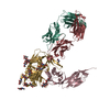
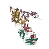
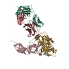
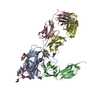
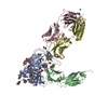
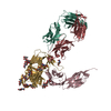
- PDB-1gc1: HIV-1 GP120 CORE COMPLEXED WITH CD4 AND A NEUTRALIZING HUMAN ANTIBODY -
+ Open data

- Basic information
| Entry | Database: PDB / ID: 1gc1 | |||||||||
|---|---|---|---|---|---|---|---|---|---|---|
| Title | HIV-1 GP120 CORE COMPLEXED WITH CD4 AND A NEUTRALIZING HUMAN ANTIBODY | |||||||||
 Components Components |
| |||||||||
 Keywords Keywords |  Viral protein/receptor/Immune system / COMPLEX (HIV ENVELOPE PROTEIN-CD4-FAB) / HIV-1 EXTERIOR ENVELOPE GP120 / T-CELL SURFACE GLYCOPROTEIN CD4 / ANTIGEN-BINDING FRAGMENT OF HUMAN IMMUNOGLOBULIN 17B / GLYCOSYLATED PROTEIN / Viral protein-receptor-Immune system COMPLEX Viral protein/receptor/Immune system / COMPLEX (HIV ENVELOPE PROTEIN-CD4-FAB) / HIV-1 EXTERIOR ENVELOPE GP120 / T-CELL SURFACE GLYCOPROTEIN CD4 / ANTIGEN-BINDING FRAGMENT OF HUMAN IMMUNOGLOBULIN 17B / GLYCOSYLATED PROTEIN / Viral protein-receptor-Immune system COMPLEX | |||||||||
| Function / homology |  Function and homology information Function and homology informationhelper T cell enhancement of adaptive immune response / interleukin-16 binding / interleukin-16 receptor activity / maintenance of protein location in cell / T cell selection / Synthesis and processing of ENV and VPU / MHC class II protein binding / evasion of host immune response / cellular response to granulocyte macrophage colony-stimulating factor stimulus / interleukin-15-mediated signaling pathway ...helper T cell enhancement of adaptive immune response / interleukin-16 binding / interleukin-16 receptor activity / maintenance of protein location in cell / T cell selection / Synthesis and processing of ENV and VPU / MHC class II protein binding / evasion of host immune response / cellular response to granulocyte macrophage colony-stimulating factor stimulus / interleukin-15-mediated signaling pathway / positive regulation of monocyte differentiation / Nef Mediated CD4 Down-regulation / Alpha-defensins / positive regulation of kinase activity / regulation of T cell activation / T cell receptor complex / extracellular matrix structural constituent / Other interleukin signaling / enzyme-linked receptor protein signaling pathway / Translocation of ZAP-70 to Immunological synapse / Phosphorylation of CD3 and TCR zeta chains / Dectin-2 family / regulation of calcium ion transport / macrophage differentiation / Generation of second messenger molecules / T cell differentiation / PD-1 signaling / positive regulation of protein kinase activity / Binding and entry of HIV virion / coreceptor activity / positive regulation of calcium-mediated signaling / positive regulation of plasma membrane raft polarization / positive regulation of receptor clustering / protein tyrosine kinase binding / positive regulation of establishment of T cell polarity / positive regulation of interleukin-2 production / virus-mediated perturbation of host defense response / T cell activation / host cell endosome membrane / actin filament organization / Vpu mediated degradation of CD4 / calcium-mediated signaling / clathrin-coated endocytic vesicle membrane / Assembly Of The HIV Virion / Budding and maturation of HIV virion / cell surface receptor protein tyrosine kinase signaling pathway / positive regulation of T cell activation / positive regulation of peptidyl-tyrosine phosphorylation / transmembrane signaling receptor activity / Cargo recognition for clathrin-mediated endocytosis / Downstream TCR signaling / virus receptor activity / signaling receptor activity / Clathrin-mediated endocytosis / MHC class II protein complex binding / clathrin-dependent endocytosis of virus by host cell / positive regulation of canonical NF-kappaB signal transduction / defense response to Gram-negative bacterium / positive regulation of MAPK cascade / adaptive immune response / positive regulation of viral entry into host cell / positive regulation of ERK1 and ERK2 cascade / cell surface receptor signaling pathway / viral protein processing / early endosome / cell adhesion / immune response / positive regulation of protein phosphorylation / symbiont entry into host cell / membrane raft / fusion of virus membrane with host plasma membrane / endoplasmic reticulum lumen / external side of plasma membrane / fusion of virus membrane with host endosome membrane / viral envelope / lipid binding / virion attachment to host cell / endoplasmic reticulum membrane / protein kinase binding / host cell plasma membrane / virion membrane / structural molecule activity / positive regulation of DNA-templated transcription / enzyme binding / signal transduction / protein homodimerization activity / zinc ion binding / membrane / identical protein binding / plasma membraneSimilarity search - Function | |||||||||
| Biological species |   Human immunodeficiency virus 1 Human immunodeficiency virus 1 Homo sapiens (human) Homo sapiens (human) | |||||||||
| Method | X-RAY DIFFRACTION / SYNCHROTRON / MOLECULAR REPLACEMENT, MIR, DENSITY MODIFICATION / Resolution: 2.5 Å | |||||||||
 Authors Authors | Kwong, P.D. / Wyatt, R. / Robinson, J. / Sweet, R.W. / Sodroski, J. / Hendrickson, W.A. | |||||||||
 Citation Citation | Journal: Nature / Year: 1998 Title: Structure of an HIV gp120 envelope glycoprotein in complex with the CD4 receptor and a neutralizing human antibody. Authors: Kwong, P.D. / Wyatt, R. / Robinson, J. / Sweet, R.W. / Sodroski, J. / Hendrickson, W.A. | |||||||||
| History |
|
- Structure visualization
| Structure viewer | Molecule: MolmilJmol/JSmol |
|---|
- Downloads & links
-Download
| PDBx/mmCIF format | 1gc1.cif.gz | 208.4 KB | Display | PDBx/mmCIF format |
|---|---|---|---|---|
| PDB format | pdb1gc1.ent.gz | 166.2 KB | Display | PDB format |
| PDBx/mmJSON format | 1gc1.json.gz | Tree view | PDBx/mmJSON format | |
| Others |  Other downloads Other downloads |
-Validation report
| Arichive directory | https://data.pdbj.org/pub/pdb/validation_reports/gc/1gc1ftp://data.pdbj.org/pub/pdb/validation_reports/gc/1gc1 | HTTPS FTP |
|---|
-Related structure data


-Links
 PDBj
PDBj


- Assembly
| Deposited unit | 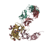
| ||||||||
|---|---|---|---|---|---|---|---|---|---|
| 1 |
| ||||||||
| Unit cell |
|
-Components
-Antibody , 3 types, 3 molecules CLH
| #2: Antibody | Mass: 20503.260 Da / Num. of mol.: 1 / Fragment: D1D2, N-TERMINAL TWO DOMAIN FRAGMENT / Mutation: S184N, I185T Source method: isolated from a genetically manipulated source Source: (gene. exp.) Homo sapiens (human) / Cell line (production host): CHO / Production host:   Cricetulus griseus (Chinese hamster) / References: UniProt: P01730 Cricetulus griseus (Chinese hamster) / References: UniProt: P01730 |
|---|---|
| #3: Antibody | Mass: 23368.818 Da / Num. of mol.: 1 / Fragment: ANTIGEN-BINDING FRAGMENT, FAB Source method: isolated from a genetically manipulated source Details: MONOCLONAL ANTIBODY 17B BINDS TO A CD4-INDUCED SITE ON GP120 Source: (gene. exp.) Homo sapiens (human)Description: EPSTEIN-BARR VIRUS IMMORTALIZED B-CELL CLONE FUSED WITH A MURINE B-CELL FUSION PARTNER Production host: Mus musculus (house mouse) |
| #4: Antibody | Mass: 24483.471 Da / Num. of mol.: 1 / Fragment: ANTIGEN-BINDING FRAGMENT, FAB Source method: isolated from a genetically manipulated source Details: MONOCLONAL ANTIBODY 17B BINDS TO A CD4-INDUCED SITE ON GP120 Source: (gene. exp.) Homo sapiens (human)Description: EPSTEIN-BARR VIRUS IMMORTALIZED B-CELL CLONE FUSED WITH A MURINE B-CELL FUSION PARTNER Production host: Mus musculus (house mouse) |
-Protein / Non-polymers , 2 types, 604 molecules G
| #1: Protein | Mass: 35429.160 Da / Num. of mol.: 1 / Fragment: CORE Mutation: (GARS) SUBSTITUTION AT THE N TERMINUS, GLY ALA GLY SUBSTITUTIONS FOR THE V1/V2 AND V3 LOOPS Source method: isolated from a genetically manipulated source Source: (gene. exp.) Human immunodeficiency virus 1 / Genus: Lentivirus / Strain: CLADE BDescription: SECRETED FROM DROSOPHILA SCHNEIDER 2 LINES UNDER CONTROL OF AN INDUCIBLE METALLOTHIONEIN PROMOTER Gene: env / Organ: OVARY / Variant: HXBC2 / Production host:  Drosophila melanogaster (fruit fly) / References: UniProt: P04578 Drosophila melanogaster (fruit fly) / References: UniProt: P04578 |
|---|---|
| #7: Water | ChemComp-HOH / WaterMass: 18.015 Da / Num. of mol.: 603 / Source method: isolated from a natural source / Formula: H2O |
-Sugars , 2 types, 11 molecules 
| #5: Polysaccharide | alpha-L-fucopyranose-(1-6)-2-acetamido-2-deoxy-beta-D-glucopyranose / Mass: 367.349 Da / Num. of mol.: 4Source method: isolated from a genetically manipulated source #6: Sugar | ChemComp-NAG / N-Acetylglucosamine Type: D-saccharide, beta linking / Mass: 221.208 Da / Num. of mol.: 7 Type: D-saccharide, beta linking / Mass: 221.208 Da / Num. of mol.: 7Source method: isolated from a genetically manipulated source Formula: C8H15NO6 |
|---|
-Experimental details
-Experiment
| Experiment | Method: X-RAY DIFFRACTION / Number of used crystals: 1 |
|---|
- Sample preparation
| Crystal | Density Matthews: 3 Å3/Da / Density % sol: 59 % | ||||||||||||||||||||||||||||||||||||||||||||||||||||||||||||||||||||||||
|---|---|---|---|---|---|---|---|---|---|---|---|---|---|---|---|---|---|---|---|---|---|---|---|---|---|---|---|---|---|---|---|---|---|---|---|---|---|---|---|---|---|---|---|---|---|---|---|---|---|---|---|---|---|---|---|---|---|---|---|---|---|---|---|---|---|---|---|---|---|---|---|---|---|
| Crystal grow | Method: vapor diffusion / pH: 7 Details: VAPOUR DIFFUSION CRYSTALLIZATION: 0.5 UL OF PROTEIN (~10MG/ML IN 350 MM NACL, 5 MM TRISCL PH 7.0) + 0.4 UL OF 0.1 M NACITRATE, 0.02 M NAHEPES, 10% ISOPROPANOL, 10.5% MONOMETHYL-PEG 5000, 0. ...Details: VAPOUR DIFFUSION CRYSTALLIZATION: 0.5 UL OF PROTEIN (~10MG/ML IN 350 MM NACL, 5 MM TRISCL PH 7.0) + 0.4 UL OF 0.1 M NACITRATE, 0.02 M NAHEPES, 10% ISOPROPANOL, 10.5% MONOMETHYL-PEG 5000, 0.0075% SEAPREP AGAROSE, PH 6.4 OVER A RESERVOIR OF 0.35 M NACL, 0.1 M NACITRATE, 0.02 M NAHEPES, 10% ISOPROPANOL, 10.5% MONOMETHYL-PEG 5000, PH 6.4, vapor diffusion PH range: 6.4-7.0 | ||||||||||||||||||||||||||||||||||||||||||||||||||||||||||||||||||||||||
| Crystal | *PLUS | ||||||||||||||||||||||||||||||||||||||||||||||||||||||||||||||||||||||||
| Crystal grow | *PLUS Temperature: 37 ℃ / pH: 6.4 / Method: vapor diffusion, hanging drop / Details: Kwong, P.D., (1999) J. Biol. Chem., 274, 4115. | ||||||||||||||||||||||||||||||||||||||||||||||||||||||||||||||||||||||||
| Components of the solutions | *PLUS
|
-Data collection
| Diffraction | Mean temperature: 100 K |
|---|---|
| Diffraction source | Source: SYNCHROTRON / Site: NSLS  / Beamline: X4A / Wavelength: 1.00614 / Beamline: X4A / Wavelength: 1.00614 |
| Detector | Type: FUJI / Detector: IMAGE PLATE / Date: Aug 1, 1996 / Details: MIRRORS |
| Radiation | Monochromator: SILICON CRYSTAL / Monochromatic (M) / Laue (L): M / Scattering type: x-ray |
| Radiation wavelength | Wavelength: 1.00614 Å / Relative weight: 1 |
| Reflection | Resolution: 2.5→20 Å / Num. obs: 37724 / % possible obs: 86 % / Observed criterion σ(I): -0.5 / Redundancy: 3 % / Rsym value: 0.093 / Net I/σ(I): 9.17 |
| Reflection shell | Resolution: 2.5→2.59 Å / Redundancy: 1.56 % / Mean I/σ(I) obs: 2.17 / Rsym value: 0.247 / % possible all: 62.8 |
| Reflection | *PLUS % possible obs: 86 % / Num. measured all: 113966 / Rmerge(I) obs: 0.093 |
- Processing
| Software |
| ||||||||||||||||||||||||||||||||||||||||||||||||||||||||||||||||||||||||||||||||
|---|---|---|---|---|---|---|---|---|---|---|---|---|---|---|---|---|---|---|---|---|---|---|---|---|---|---|---|---|---|---|---|---|---|---|---|---|---|---|---|---|---|---|---|---|---|---|---|---|---|---|---|---|---|---|---|---|---|---|---|---|---|---|---|---|---|---|---|---|---|---|---|---|---|---|---|---|---|---|---|---|---|
| Refinement | Method to determine structure: MOLECULAR REPLACEMENT, MIR, DENSITY MODIFICATION Starting model: PDB ENTRIES 1HIL, 1CDH AND 3CD4 Resolution: 2.5→5 Å / Data cutoff high absF: 100000 / Data cutoff low absF: 0.1 / Isotropic thermal model: RESTRAINED / Cross valid method: THROUGHOUT / σ(F): 2
| ||||||||||||||||||||||||||||||||||||||||||||||||||||||||||||||||||||||||||||||||
| Displacement parameters | Biso mean: 21 Å2
| ||||||||||||||||||||||||||||||||||||||||||||||||||||||||||||||||||||||||||||||||
| Refine analyze | Luzzati d res low obs: 5 Å | ||||||||||||||||||||||||||||||||||||||||||||||||||||||||||||||||||||||||||||||||
| Refinement step | Cycle: LAST / Resolution: 2.5→5 Å
| ||||||||||||||||||||||||||||||||||||||||||||||||||||||||||||||||||||||||||||||||
| Refine LS restraints |
| ||||||||||||||||||||||||||||||||||||||||||||||||||||||||||||||||||||||||||||||||
| LS refinement shell | Resolution: 2.5→2.58 Å / Total num. of bins used: 10
| ||||||||||||||||||||||||||||||||||||||||||||||||||||||||||||||||||||||||||||||||
| Xplor file |
| ||||||||||||||||||||||||||||||||||||||||||||||||||||||||||||||||||||||||||||||||
| Software | *PLUS Name: X-PLOR / Version: 3.8 / Classification: refinement | ||||||||||||||||||||||||||||||||||||||||||||||||||||||||||||||||||||||||||||||||
| Refinement | *PLUS Rfactor Rfree: 0.3026 | ||||||||||||||||||||||||||||||||||||||||||||||||||||||||||||||||||||||||||||||||
| Solvent computation | *PLUS | ||||||||||||||||||||||||||||||||||||||||||||||||||||||||||||||||||||||||||||||||
| Displacement parameters | *PLUS |
