Movie
Movie Controller

[English] 日本語
 Yorodumi
- EMDB-1900: Cryo-EM map of the SPP1 bacteriophage gp19.1-gp21(1-552) complex -
+ Open data

- Basic information
| Entry | Database: EMDB / ID: EMD-1900 | |||||||||
|---|---|---|---|---|---|---|---|---|---|---|
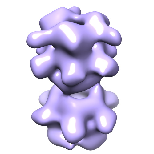
| Title | Cryo-EM map of the SPP1 bacteriophage gp19.1-gp21(1-552) complex | |||||||||
 Map data Map data | CCP4 Map file of Bacteriophage spp1 Dit-Tal complex | |||||||||
 Sample Sample |
| |||||||||
 Keywords Keywords | Bacteriophage / SPP1 / Tail tip / gp19.1 / Dit / gp21 / Tal / tail adsorption apparatus / infection mechanism | |||||||||
| Biological species |  unidentified phage (virus) unidentified phage (virus) | |||||||||
| Method | single particle reconstruction / cryo EM / Resolution: 26.0 Å | |||||||||
 Authors Authors | Goulet A / Lai-Kee-Him J / Veesler D / Auzat I / Robin G / Shepherd DA / Ashcroft AE / Richard E / Lichiere J / Tavares P ...Goulet A / Lai-Kee-Him J / Veesler D / Auzat I / Robin G / Shepherd DA / Ashcroft AE / Richard E / Lichiere J / Tavares P / Cambillau C / Bron P | |||||||||
 Citation Citation | Journal: J Biol Chem / Year: 2011 Title: The opening of the SPP1 bacteriophage tail, a prevalent mechanism in Gram-positive-infecting siphophages. Authors: Adeline Goulet / Joséphine Lai-Kee-Him / David Veesler / Isabelle Auzat / Gautier Robin / Dale A Shepherd / Alison E Ashcroft / Eric Richard / Julie Lichière / Paulo Tavares / Christian ...Authors: Adeline Goulet / Joséphine Lai-Kee-Him / David Veesler / Isabelle Auzat / Gautier Robin / Dale A Shepherd / Alison E Ashcroft / Eric Richard / Julie Lichière / Paulo Tavares / Christian Cambillau / Patrick Bron /  Abstract: The SPP1 siphophage uses its long non-contractile tail and tail tip to recognize and infect the Gram-positive bacterium Bacillus subtilis. The tail-end cap and its attached tip are the critical ...The SPP1 siphophage uses its long non-contractile tail and tail tip to recognize and infect the Gram-positive bacterium Bacillus subtilis. The tail-end cap and its attached tip are the critical components for host recognition and opening of the tail tube for genome exit. In the present work, we determined the cryo-electron microscopic (cryo-EM) structure of a complex formed by the cap protein gp19.1 (Dit) and the N terminus of the downstream protein of gp19.1 in the SPP1 genome, gp21(1-552) (Tal). This complex assembles two back-to-back stacked gp19.1 ring hexamers, interacting loosely, and two gp21(1-552) trimers interacting with gp19.1 at both ends of the stack. Remarkably, one gp21(1-552) trimer displays a "closed" conformation, whereas the second is "open" delineating a central channel. The two conformational states dock nicely into the EM map of the SPP1 cap domain, respectively, before and after DNA release. Moreover, the open/closed conformations of gp19.1-gp21(1-552) are consistent with the structures of the corresponding proteins in the siphophage p2 baseplate, where the Tal protein (ORF16) attached to the ring of Dit (ORF15) was also found to adopt these two conformations. Therefore, the present contribution allowed us to revisit the SPP1 tail distal-end architectural organization. Considering the sequence conservation among Dit and the N-terminal region of Tal-like proteins in Gram-positive-infecting Siphoviridae, it also reveals the Tal opening mechanism as a hallmark of siphophages probably involved in the generation of the firing signal initiating the cascade of events that lead to phage DNA release in vivo. | |||||||||
| History |
|
- Structure visualization
| Movie |
 Movie viewer Movie viewer |
|---|---|
| Structure viewer | EM map: SurfViewMolmilJmol/JSmol |
| Supplemental images |
- Downloads & links
-EMDB archive
| Map data | emd_1900.map.gz | 3 MB | EMDB map data format | |
|---|---|---|---|---|
| Header (meta data) | emd-1900-v30.xmlemd-1900.xml | 11 KB 11 KB | Display Display | EMDB header |
| Images |  emd-1900.jpg emd-1900.jpg | 75.2 KB | ||
| Archive directory |  http://ftp.pdbj.org/pub/emdb/structures/EMD-1900ftp://ftp.pdbj.org/pub/emdb/structures/EMD-1900 http://ftp.pdbj.org/pub/emdb/structures/EMD-1900ftp://ftp.pdbj.org/pub/emdb/structures/EMD-1900 | HTTPS FTP |
-Validation report
| Summary document | emd_1900_validation.pdf.gz | 210.6 KB | Display | EMDB validaton report |
|---|---|---|---|---|
| Full document | emd_1900_full_validation.pdf.gz | 209.7 KB | Display | |
| Data in XML | emd_1900_validation.xml.gz | 5.2 KB | Display | |
| Arichive directory | https://ftp.pdbj.org/pub/emdb/validation_reports/EMD-1900ftp://ftp.pdbj.org/pub/emdb/validation_reports/EMD-1900 | HTTPS FTP |
-Related structure data
| Similar structure data |
|---|


-Links
| EMDB pages | EMDB (EBI/PDBe) / EMDataResource |
|---|
-Map
| File | Download / File: emd_1900.map.gz / Format: CCP4 / Size: 3.7 MB / Type: IMAGE STORED AS FLOATING POINT NUMBER (4 BYTES) | ||||||||||||||||||||||||||||||||||||||||||||||||||||||||||||||||||||
|---|---|---|---|---|---|---|---|---|---|---|---|---|---|---|---|---|---|---|---|---|---|---|---|---|---|---|---|---|---|---|---|---|---|---|---|---|---|---|---|---|---|---|---|---|---|---|---|---|---|---|---|---|---|---|---|---|---|---|---|---|---|---|---|---|---|---|---|---|---|
| Annotation | CCP4 Map file of Bacteriophage spp1 Dit-Tal complex | ||||||||||||||||||||||||||||||||||||||||||||||||||||||||||||||||||||
| Voxel size | X=Y=Z: 4.38 Å | ||||||||||||||||||||||||||||||||||||||||||||||||||||||||||||||||||||
| Density |
| ||||||||||||||||||||||||||||||||||||||||||||||||||||||||||||||||||||
| Symmetry | Space group: 1 | ||||||||||||||||||||||||||||||||||||||||||||||||||||||||||||||||||||
| Details | EMDB XML:
CCP4 map header:
| ||||||||||||||||||||||||||||||||||||||||||||||||||||||||||||||||||||
-Supplemental data
- Sample components
-Entire : SPP1 bacteriophage gp19.1-gp21(1-552) complex
| Entire | Name: SPP1 bacteriophage gp19.1-gp21(1-552) complex |
|---|---|
| Components |
|
-Supramolecule #1000: SPP1 bacteriophage gp19.1-gp21(1-552) complex
| Supramolecule | Name: SPP1 bacteriophage gp19.1-gp21(1-552) complex / type: sample / ID: 1000 Oligomeric state: This complex assembles two back-to- back stacked gp19.1 ring hexamers, interacting loosely, and two gp21(1-552) trimers interacting with gp19.1 at both ends of the stack Number unique components: 2 |
|---|---|
| Molecular weight | Experimental: 730 KDa / Method: SEC, MALS, RI, UV, QELS |
-Macromolecule #1: gp19.1
| Macromolecule | Name: gp19.1 / type: protein_or_peptide / ID: 1 / Name.synonym: Dit / Recombinant expression: Yes |
|---|---|
| Source (natural) | Organism: unidentified phage (virus) / synonym: Dit |
-Macromolecule #2: gp21(1-552)
| Macromolecule | Name: gp21(1-552) / type: protein_or_peptide / ID: 2 / Name.synonym: Tal / Recombinant expression: Yes |
|---|---|
| Source (natural) | Organism: unidentified phage (virus) / synonym: Tal |
-Experimental details
-Structure determination
| Method | cryo EM |
|---|---|
 Processing Processing | single particle reconstruction |
| Aggregation state | particle |
-Sample preparation
| Concentration | 0.07 mg/mL |
|---|---|
| Buffer | pH: 7.5 / Details: 10 mM HEPES, 150 mM NaCl |
| Grid | Details: Quantifoil R 2/2 grids |
| Vitrification | Cryogen name: ETHANE / Chamber humidity: 90 % / Chamber temperature: 102.15 K / Instrument: GATAN CRYOPLUNGE 3 / Details: Vitrification instrument: Cryoplunge CP3 gatan / Method: Blot for 2s, both sides |
- Electron microscopy
| Microscope | JEOL 2200FS |
|---|---|
| Temperature | Average: 98.15 K |
| Alignment procedure | Legacy - Astigmatism: Objective lens astigmatism was corrected at 200,000 times magnification |
| Specialist optics | Energy filter - Name: Omega JEOL / Energy filter - Lower energy threshold: 0.0 eV / Energy filter - Upper energy threshold: 20.0 eV |
| Details | Images recorded on a JEOL 2200FS |
| Image recording | Category: FILM / Film or detector model: KODAK SO-163 FILM / Digitization - Scanner: NIKON SUPER COOLSCAN 9000 / Digitization - Sampling interval: 10 µm / Number real images: 100 / Average electron dose: 20 e/Å2 |
| Electron beam | Acceleration voltage: 200 kV / Electron source:  FIELD EMISSION GUN FIELD EMISSION GUN |
| Electron optics | Calibrated magnification: 45591 / Illumination mode: FLOOD BEAM / Imaging mode: BRIGHT FIELD / Cs: 2.0 mm / Nominal defocus max: 2.5 µm / Nominal defocus min: 1.4 µm / Nominal magnification: 50000 |
| Sample stage | Specimen holder: Eucentric / Specimen holder model: SIDE ENTRY, EUCENTRIC |
-Image processing
| CTF correction | Details: Each particle |
|---|---|
| Final reconstruction | Applied symmetry - Point group: D3 (2x3 fold dihedral) / Algorithm: OTHER / Resolution.type: BY AUTHOR / Resolution: 26.0 Å / Resolution method: FSC 0.5 CUT-OFF / Software - Name: IMAGIC / Number images used: 15171 |
-Atomic model buiding 1
| Initial model | PDB ID: |
|---|---|
| Details | The initial fit was done by manual docking followed by refinement using the (fit in map) Chimera plugging |
| Refinement | Space: REAL / Protocol: RIGID BODY FIT |