ムービー
ムービー コントローラー


+ データを開く

- 基本情報
基本情報
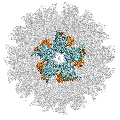
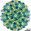
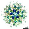
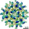
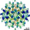
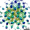
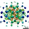
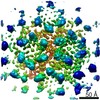
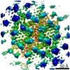
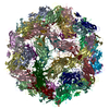
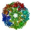
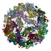
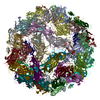
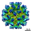
| 登録情報 | データベース: EMDB / ID: EMD-12485 | |||||||||||||||
|---|---|---|---|---|---|---|---|---|---|---|---|---|---|---|---|---|
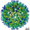
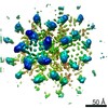
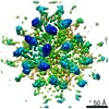
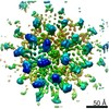
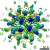
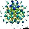
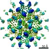
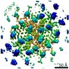
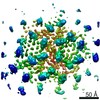
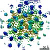
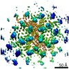
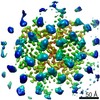
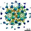
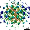
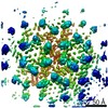
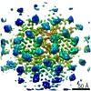
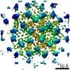
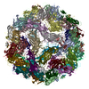
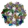
| タイトル | Structure of the mature RSV CA lattice: T=1 CA icosahedron | |||||||||||||||
 マップデータ マップデータ | RSV CA T=1 particle | |||||||||||||||
 試料 試料 |
| |||||||||||||||
| 機能・相同性 |  機能・相同性情報 機能・相同性情報host cell nucleoplasm / viral procapsid maturation / host cell nucleolus /  加水分解酵素; プロテアーゼ; ペプチド結合加水分解酵素; アスパラギン酸プロテアーゼ / カプシド / structural constituent of virion / nucleic acid binding / aspartic-type endopeptidase activity / host cell plasma membrane / タンパク質分解 ...host cell nucleoplasm / viral procapsid maturation / host cell nucleolus / 加水分解酵素; プロテアーゼ; ペプチド結合加水分解酵素; アスパラギン酸プロテアーゼ / カプシド / structural constituent of virion / nucleic acid binding / aspartic-type endopeptidase activity / host cell plasma membrane / タンパク質分解 / zinc ion binding / 生体膜 加水分解酵素; プロテアーゼ; ペプチド結合加水分解酵素; アスパラギン酸プロテアーゼ / カプシド / structural constituent of virion / nucleic acid binding / aspartic-type endopeptidase activity / host cell plasma membrane / タンパク質分解 ...host cell nucleoplasm / viral procapsid maturation / host cell nucleolus / 加水分解酵素; プロテアーゼ; ペプチド結合加水分解酵素; アスパラギン酸プロテアーゼ / カプシド / structural constituent of virion / nucleic acid binding / aspartic-type endopeptidase activity / host cell plasma membrane / タンパク質分解 / zinc ion binding / 生体膜類似検索 - 分子機能 | |||||||||||||||
| 生物種 |  Rous sarcoma virus (strain Prague C) (ラウス肉腫ウイルス) / Rous sarcoma virus - Prague C (ラウス肉腫ウイルス) Rous sarcoma virus (strain Prague C) (ラウス肉腫ウイルス) / Rous sarcoma virus - Prague C (ラウス肉腫ウイルス) | |||||||||||||||
| 手法 | 単粒子再構成法 / クライオ電子顕微鏡法 / 解像度: 3.1 Å | |||||||||||||||
 データ登録者 データ登録者 | Obr M / Ricana CL / Nikulin N / Feathers J-PR / Klanschnig M / Thader A / Johnson MC / Vogt VM / Schur FKM / Dick RA | |||||||||||||||
| 資金援助 |  オーストリア, オーストリア,  米国, 4件 米国, 4件
| |||||||||||||||
 引用 引用 | ジャーナル: Nat Commun / 年: 2021 タイトル: Structure of the mature Rous sarcoma virus lattice reveals a role for IP6 in the formation of the capsid hexamer. 著者: Martin Obr / Clifton L Ricana / Nadia Nikulin / Jon-Philip R Feathers / Marco Klanschnig / Andreas Thader / Marc C Johnson / Volker M Vogt / Florian K M Schur / Robert A Dick / 要旨: Inositol hexakisphosphate (IP6) is an assembly cofactor for HIV-1. We report here that IP6 is also used for assembly of Rous sarcoma virus (RSV), a retrovirus from a different genus. IP6 is ~100-fold ...Inositol hexakisphosphate (IP6) is an assembly cofactor for HIV-1. We report here that IP6 is also used for assembly of Rous sarcoma virus (RSV), a retrovirus from a different genus. IP6 is ~100-fold more potent at promoting RSV mature capsid protein (CA) assembly than observed for HIV-1 and removal of IP6 in cells reduces infectivity by 100-fold. Here, visualized by cryo-electron tomography and subtomogram averaging, mature capsid-like particles show an IP6-like density in the CA hexamer, coordinated by rings of six lysines and six arginines. Phosphate and IP6 have opposing effects on CA in vitro assembly, inducing formation of T = 1 icosahedrons and tubes, respectively, implying that phosphate promotes pentamer and IP6 hexamer formation. Subtomogram averaging and classification optimized for analysis of pleomorphic retrovirus particles reveal that the heterogeneity of mature RSV CA polyhedrons results from an unexpected, intrinsic CA hexamer flexibility. In contrast, the CA pentamer forms rigid units organizing the local architecture. These different features of hexamers and pentamers determine the structural mechanism to form CA polyhedrons of variable shape in mature RSV particles. | |||||||||||||||
| 履歴 |
|
- 構造の表示
構造の表示
| ムービー |
ムービービューア |
|---|---|
| 構造ビューア | EMマップ: SurfViewMolmilJmol/JSmol |
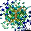
| 添付画像 |
- ダウンロードとリンク
-EMDBアーカイブ
| マップデータ | emd_12485.map.gz | 27.3 MB | EMDBマップデータ形式 | |
|---|---|---|---|---|
| ヘッダ (付随情報) | emd-12485-v30.xmlemd-12485.xml | 16.8 KB 16.8 KB | 表示 表示 | EMDBヘッダ |
| 画像 |  emd_12485.png emd_12485.png | 248.4 KB | ||
| アーカイブディレクトリ |  http://ftp.pdbj.org/pub/emdb/structures/EMD-12485ftp://ftp.pdbj.org/pub/emdb/structures/EMD-12485 http://ftp.pdbj.org/pub/emdb/structures/EMD-12485ftp://ftp.pdbj.org/pub/emdb/structures/EMD-12485 | HTTPS FTP |
-関連構造データ
| 関連構造データ | 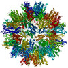 7no0MC 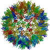 7no1C 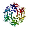 7no2C  7no3C  7no4C  7no5C  7no6C 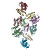 7no7C 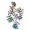 7no8C 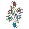 7no9C 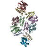 7noaC 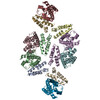 7nobC 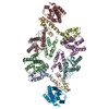 7nocC 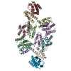 7nodC 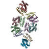 7noeC  7nofC 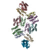 7nogC 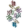 7nohC 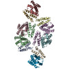 7noiC 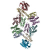 7nojC 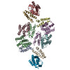 7nokC 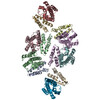 7nolC 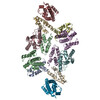 7nomC 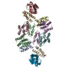 7nonC 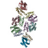 7nooC 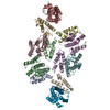 7nopC 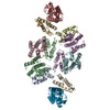 7noqC M: このマップから作成された原子モデル C: 同じ文献を引用 ( |
|---|---|
| 類似構造データ |
-リンク
| EMDBのページ | EMDB (EBI/PDBe) / EMDataResource |
|---|
-マップ
| ファイル | ダウンロード / ファイル: emd_12485.map.gz / 形式: CCP4 / 大きさ: 343 MB / タイプ: IMAGE STORED AS FLOATING POINT NUMBER (4 BYTES) | ||||||||||||||||||||||||||||||||||||||||||||||||||||||||||||
|---|---|---|---|---|---|---|---|---|---|---|---|---|---|---|---|---|---|---|---|---|---|---|---|---|---|---|---|---|---|---|---|---|---|---|---|---|---|---|---|---|---|---|---|---|---|---|---|---|---|---|---|---|---|---|---|---|---|---|---|---|---|
| 注釈 | RSV CA T=1 particle | ||||||||||||||||||||||||||||||||||||||||||||||||||||||||||||
| ボクセルのサイズ | X=Y=Z: 1.24 Å | ||||||||||||||||||||||||||||||||||||||||||||||||||||||||||||
| 密度 |
| ||||||||||||||||||||||||||||||||||||||||||||||||||||||||||||
| 対称性 | 空間群: 1 | ||||||||||||||||||||||||||||||||||||||||||||||||||||||||||||
| 詳細 | EMDB XML:
CCP4マップ ヘッダ情報:
| ||||||||||||||||||||||||||||||||||||||||||||||||||||||||||||
-添付データ
- 試料の構成要素
-全体 : Rous sarcoma virus - Prague C
| 全体 | 名称: Rous sarcoma virus - Prague C (ラウス肉腫ウイルス) |
|---|---|
| 要素 |
|
-超分子 #1: Rous sarcoma virus - Prague C
| 超分子 | 名称: Rous sarcoma virus - Prague C / タイプ: virus / ID: 1 / 親要素: 0 / 含まれる分子: all / NCBI-ID: 11888 / 生物種: Rous sarcoma virus - Prague C / ウイルスタイプ: VIRUS-LIKE PARTICLE / ウイルス・単離状態: OTHER / ウイルス・エンベロープ: No / ウイルス・中空状態: Yes |
|---|---|
| Host system | 生物種:  Escherichia coli BL21(DE3) (大腸菌) Escherichia coli BL21(DE3) (大腸菌) |
| ウイルス殻 | Shell ID: 1 / 名称: T=1 icosahedron |
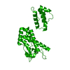
-分子 #1: Capsid protein p27, alternate cleaved 1
| 分子 | 名称: Capsid protein p27, alternate cleaved 1 / タイプ: protein_or_peptide / ID: 1 / コピー数: 1 / 光学異性体: LEVO |
|---|---|
| 由来(天然) | 生物種: Rous sarcoma virus (strain Prague C) (ラウス肉腫ウイルス) 株: Prague C |
| 分子量 | 理論値: 24.773594 KDa |
| 組換発現 | 生物種: Escherichia coli BL21(DE3) (大腸菌) |
| 配列 | 文字列: PVVIKTEGPA WTPLEPKLIT RLADTVRTKG LRSPITMAEV EALMSSPLLP HDVTNLMRVI LGPAPYALWM DAWGVQLQTV IAAATRDPR HPANGQGRGE RTNLNRLKGL ADGMVGNPQG QAALLRPGEL VAITASALQA FREVARLAEP AGPWADIMQG P SESFVDFA ...文字列: PVVIKTEGPA WTPLEPKLIT RLADTVRTKG LRSPITMAEV EALMSSPLLP HDVTNLMRVI LGPAPYALWM DAWGVQLQTV IAAATRDPR HPANGQGRGE RTNLNRLKGL ADGMVGNPQG QAALLRPGEL VAITASALQA FREVARLAEP AGPWADIMQG P SESFVDFA NRLIKAVEGS DLPPSARAPV IIDCFRQKSQ PDIQQLIRTA PSTLTTPGEI IKYVLDRQKT A |
-実験情報
-構造解析
| 手法 | クライオ電子顕微鏡法 |
|---|---|
 解析 解析 | 単粒子再構成法 |
| 試料の集合状態 | particle |
-試料調製
| 緩衝液 | pH: 8 / 構成要素 - 濃度: 1.0 M / 構成要素 - 式: NaPi / 構成要素 - 名称: sodium phosphate |
|---|---|
| グリッド | モデル: C-flat-2/2 / 材質: COPPER / メッシュ: 300 / 支持フィルム - 材質: CARBON / 支持フィルム - トポロジー: HOLEY / 前処理 - タイプ: GLOW DISCHARGE / 前処理 - 雰囲気: AIR |
| 凍結 | 凍結剤: ETHANE / チャンバー内湿度: 95 % / チャンバー内温度: 277 K / 装置: FEI VITROBOT MARK IV / 詳細: blot time= 2.5 s blot force= 0. |
- 電子顕微鏡法
| 顕微鏡 | FEI TECNAI ARCTICA |
|---|---|
| 電子線 | 加速電圧: 200 kV / 電子線源: FIELD EMISSION GUN |
| 電子光学系 | C2レンズ絞り径: 50.0 µm / 照射モード: FLOOD BEAM / 撮影モード: BRIGHT FIELDBright-field microscopy / Cs: 2.7 mm / 最大 デフォーカス(公称値): 1.5 µm / 最小 デフォーカス(公称値): 0.8 µm / 倍率(公称値): 63000 |
| 特殊光学系 | エネルギーフィルター - 名称: GIF Bioquantum / エネルギーフィルター - スリット幅: 20 eV |
| 試料ステージ | 試料ホルダーモデル: FEI TITAN KRIOS AUTOGRID HOLDER ホルダー冷却材: NITROGEN |
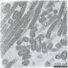
| 撮影 | フィルム・検出器のモデル: GATAN K3 BIOQUANTUM (6k x 4k) デジタル化 - サイズ - 横: 5760 pixel / デジタル化 - サイズ - 縦: 4092 pixel / 平均露光時間: 3.0 sec. / 平均電子線量: 50.0 e/Å2 |
| 実験機器 |  モデル: Talos Arctica / 画像提供: FEI Company |
-画像解析
| 粒子像選択 | 選択した数: 150599 詳細: 2394 micrographs were taken, from which 374 particles were manually picked and 2D classified to generate templates for auto-picking. Two rounds of auto-picking and 2D classification resulted ...詳細: 2394 micrographs were taken, from which 374 particles were manually picked and 2D classified to generate templates for auto-picking. Two rounds of auto-picking and 2D classification resulted in 150599 extracted particles. |
|---|---|
| CTF補正 | ソフトウェア: (名称: Gctf, RELION) 詳細: CTF estimation and correction was performed using GCTF in the RELION wrapper |
| 初期モデル | モデルのタイプ: EMDB MAP EMDB ID: 詳細: Map was low pass filtered to 60 Angstrom^-1 |
| 初期 角度割当 | タイプ: MAXIMUM LIKELIHOOD / ソフトウェア - 名称: RELION |
| 最終 3次元分類 | ソフトウェア - 名称: RELION 詳細: 2D and 3D classification, CTF refinement and Bayesian particle polishing |
| 最終 角度割当 | タイプ: MAXIMUM LIKELIHOOD / ソフトウェア - 名称: RELION |
| 最終 再構成 | 想定した対称性 - 点群: I (正20面体型対称) / 解像度のタイプ: BY AUTHOR / 解像度: 3.1 Å / 解像度の算出法: FSC 0.143 CUT-OFF / ソフトウェア - 名称: RELION / 使用した粒子像数: 20498 |
-原子モデル構築 1
| 初期モデル | PDB ID: |
|---|---|
| 詳細 | The CANTD and CACTD of one CA monomer of pdb 3TIR were independently placed into the EM-density using the rigid body fitting option in UCSF Chimera. Subsequently the linker connecting the two CA domains was joined in Coot. To account for the different monomer-monomer interactions in the RSV CA pentamer, the monomers were replicated according to the inherent 5-fold symmetry of the map and an additional ring of CACTDs was rigid-body fitted into the EM-densities of the surrounding CA pentamers. A map segment (defined by a mask extending 3 Angstrom around the rigid body fitted model) was extracted, and real-space coordinate refinement against the EM-density was performed using Phenix. This was iterated with manual model building in Coot, similar as described previously. In brief, secondary structure restraints and non-crystallographic symmetry (NCS) restraints were applied throughout all refinements. Each Phenix iteration consisted of 5 macro cycles, in which simulated annealing was performed in every macro cycle. Atomic displacement parameter (ADP) refinement was per formed at the end of each iteration. |
| 精密化 | 空間: REAL / プロトコル: AB INITIO MODEL |
| 得られたモデル | PDB-7no0: |