 Movie
Movie Controller

[English] 日本語
 Yorodumi
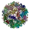
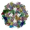
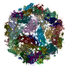
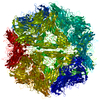
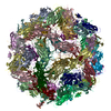
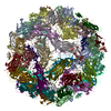
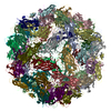
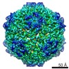
- PDB-3jci: 2.9 Angstrom Resolution Cryo-EM 3-D Reconstruction of Close-packe... -
+ Open data

- Basic information
| Entry | Database: PDB / ID: 3jci | ||||||
|---|---|---|---|---|---|---|---|
| Title | 2.9 Angstrom Resolution Cryo-EM 3-D Reconstruction of Close-packed PCV2 Virus-like Particles | ||||||
 Components Components | Capsid protein | ||||||
 Keywords Keywords | VIRUS LIKE PARTICLE / de novo initial model / consensus criterion / gold-standard FSC / true FSC / cross-validation | ||||||
| Function / homology |  Function and homology information Function and homology informationviral capsid assembly / T=1 icosahedral viral capsid / endocytosis involved in viral entry into host cell / viral penetration into host nucleus / host cell nucleus / virion attachment to host cell / nucleus Similarity search - Function | ||||||
| Biological species |  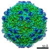 Porcine circovirus-2 Porcine circovirus-2 | ||||||
| Method | ELECTRON MICROSCOPY / single particle reconstruction / cryo EM / Resolution: 2.9 Å | ||||||
 Authors Authors | Liu, Z. / Guo, F. / Wang, F. / Li, T.C. / Jiang, W. | ||||||
 Citation Citation | Journal: Structure / Year: 2016 Title: 2.9 Å Resolution Cryo-EM 3D Reconstruction of Close-Packed Virus Particles. Authors: Zheng Liu / Fei Guo / Feng Wang / Tian-Cheng Li / Wen Jiang /   Abstract: Single-particle cryoelectron microscopy typically discards close-packed particle images as unusable data. Here, we report an image processing strategy and case study of obtaining near-atomic ...Single-particle cryoelectron microscopy typically discards close-packed particle images as unusable data. Here, we report an image processing strategy and case study of obtaining near-atomic resolution 3D reconstructions from close-packed particles. Multiple independent de novo initial models were constructed to determine and cross-validate the particle parameters. The particles with consistent views were further refined including not only Euler angles and center positions but also defocus, astigmatism, beam tilt, and overall and anisotropic magnification. We demonstrated this strategy with a 2.9 Å resolution reconstruction of a 1.67 MDa virus-like particle of a circovirus, PCV2, recorded on 86 photographic films. The map resolution was further validated with a phase-randomization test and local resolution assessment, and the atomic model was validated with MolProbity and EMRinger. Close-packed virus particles were thus shown not only to be useful for high-resolution 3D reconstructions but also to allow data collection at significantly improved throughput for near-atomic resolution reconstructions. | ||||||
| History |
|
- Structure visualization
| Movie |
Movie viewer |
|---|---|
| Structure viewer | Molecule: MolmilJmol/JSmol |
- Downloads & links
-Download
| PDBx/mmCIF format | 3jci.cif.gz | 51.8 KB | Display | PDBx/mmCIF format |
|---|---|---|---|---|
| PDB format | pdb3jci.ent.gz | 35.8 KB | Display | PDB format |
| PDBx/mmJSON format | 3jci.json.gz | Tree view | PDBx/mmJSON format | |
| Others |  Other downloads Other downloads |
-Validation report
| Summary document | 3jci_validation.pdf.gz | 769 KB | Display | wwPDB validaton report |
|---|---|---|---|---|
| Full document | 3jci_full_validation.pdf.gz | 770.4 KB | Display | |
| Data in XML | 3jci_validation.xml.gz | 14.7 KB | Display | |
| Data in CIF | 3jci_validation.cif.gz | 19.1 KB | Display | |
| Arichive directory | https://data.pdbj.org/pub/pdb/validation_reports/jc/3jciftp://data.pdbj.org/pub/pdb/validation_reports/jc/3jci | HTTPS FTP |
-Related structure data
| Related structure data |  6555MC M: map data used to model this data C: citing same article ( |
|---|---|
| Similar structure data |
-Links
 PDBj
PDBj
- Assembly
| Deposited unit | 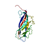
|
|---|---|
| 1 | x 60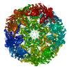
|
| 2 |
|
| 3 | x 5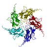
|
| 4 | x 6
|
| 5 | 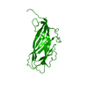
|
| Symmetry | Point symmetry: (Schoenflies symbol: I (icosahedral)) |
-Components
| #1: Protein | Mass: 21999.758 Da / Num. of mol.: 1 / Fragment: UNP residues 42-231 / Source method: isolated from a natural source / Source: (natural) Porcine circovirus-2 / References: UniProt: B8Y5Y9, UniProt: Q8B980*PLUS |
|---|
-Experimental details
-Experiment
| Experiment | Method: ELECTRON MICROSCOPY |
|---|---|
| EM experiment | Aggregation state: PARTICLE / 3D reconstruction method: single particle reconstruction |
- Sample preparation
| Component |
| ||||||||||||
|---|---|---|---|---|---|---|---|---|---|---|---|---|---|
| Molecular weight | Value: 1.67 MDa / Experimental value: NO | ||||||||||||
| Details of virus | Empty: YES / Enveloped: NO / Host category: VERTEBRATES / Isolate: STRAIN / Type: VIRUS-LIKE PARTICLE | ||||||||||||
| Natural host | Organism: Sus scrofa | ||||||||||||
| Buffer solution | Name: PBS / pH: 7.4 / Details: PBS | ||||||||||||
| Specimen | Conc.: 3 mg/ml / Embedding applied: NO / Shadowing applied: NO / Staining applied: NO / Vitrification applied: YES | ||||||||||||
| Specimen support | Details: 400-mesh holey carbon grids (1.2/1.3 C-flat, Protochips) | ||||||||||||
| Vitrification | Instrument: GATAN CRYOPLUNGE 3 / Cryogen name: ETHANE / Temp: 85 K / Humidity: 90 % Details: Blot for 5 seconds before plunging into liquid ethane (GATAN CRYOPLUNGE 3). Method: Blot for 5 seconds before plunging |
- Electron microscopy imaging
| Experimental equipment |  Model: Titan Krios / Image courtesy: FEI Company |
|---|---|
| Microscopy | Model: FEI TITAN KRIOS / Date: Feb 22, 2011 |
| Electron gun | Electron source:  FIELD EMISSION GUN / Accelerating voltage: 300 kV / Illumination mode: FLOOD BEAM FIELD EMISSION GUN / Accelerating voltage: 300 kV / Illumination mode: FLOOD BEAM |
| Electron lens | Mode: BRIGHT FIELD / Nominal magnification: 59000 X / Calibrated magnification: 587963 X / Nominal defocus max: 2500 nm / Nominal defocus min: 200 nm / Cs: 2.7 mm Astigmatism: Objective lens astigmatism was corrected at 250,000 magnification using quadrupole stigmator. |
| Specimen holder | Specimen holder model: FEI TITAN KRIOS AUTOGRID HOLDER / Temperature: 90 K / Temperature (max): 100 K / Temperature (min): 80 K |
| Image recording | Electron dose: 25 e/Å2 / Film or detector model: KODAK SO-163 FILM |
| Image scans | Num. digital images: 141 |
- Processing
| EM software |
| ||||||||||||||||||||||||
|---|---|---|---|---|---|---|---|---|---|---|---|---|---|---|---|---|---|---|---|---|---|---|---|---|---|
| CTF correction | Details: Each particle | ||||||||||||||||||||||||
| Symmetry | Point symmetry: I (icosahedral) | ||||||||||||||||||||||||
| 3D reconstruction | Method: Projection matching / Resolution: 2.9 Å / Resolution method: FSC 0.143 CUT-OFF / Num. of particles: 50352 / Nominal pixel size: 1.1 Å / Actual pixel size: 1.08 Å Details: For 3D reconstruction, whole datasets were divided into even and odd halves and the initial de novo models and subsequent iterative refinements were all independently performed for each half ...Details: For 3D reconstruction, whole datasets were divided into even and odd halves and the initial de novo models and subsequent iterative refinements were all independently performed for each half dataset. Particles were selected from scanned micrograph images using e2boxer.py in EMAN2. The TEM instrument contrast transfer function parameters were determined automatically using fitctf2.py in JSPR and were then visually validated using the EMAN ctfit program. The datasets were then divided into two subsets (even and odd) and processed completely independently, including both de novo initial models and refinements. The images were first binned 4x to obtain initial models and particle parameters assuming icosahedral symmetry. De novo initial models were built using the random model approach. Random subsets of particles were assigned random initial orientations and iteratively refined until convergence. Multi-model competitive refinements were used to choose the winning model (with most assigned particles) as corrective initial models for subsequent refinement. Particles with inconsistent/unstable view parameters in the initial refinements were excluded in further image processing. The orientation and center parameters were then transferred to the un-binned images for high-resolution refinements which included Simplex method-based orientation/center optimization and grid search-based refinement of defocus, astigmatism, beam tilt, and overall and anisotropic magnification of the images. All image refinement and reconstructions were performed with JSPR software that was built on EMAN2 and EMAN library functions and programs. (Single particle details: The particles were selected using the e2boxer.py program in EMAN2. CTF parameters were determined using fitctf2.py in JSPR.) (Single particle--Applied symmetry: I) Symmetry type: POINT | ||||||||||||||||||||||||
| Displacement parameters | Biso max: 77.94 Å2 / Biso mean: 23.672 Å2 / Biso min: 10.28 Å2 | ||||||||||||||||||||||||
| Refinement step | Cycle: LAST
| ||||||||||||||||||||||||
| Refine LS restraints |
|
