Movie
Movie Controller


+ Open data

- Basic information
| Entry | Database: PDB / ID: 7no0 | |||||||||||||||
|---|---|---|---|---|---|---|---|---|---|---|---|---|---|---|---|---|
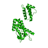
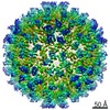
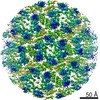
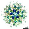
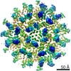
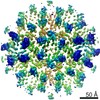
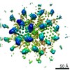
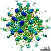
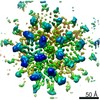
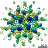
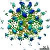
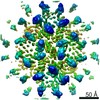
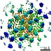
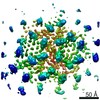
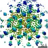
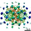
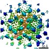
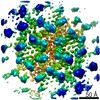
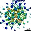
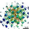
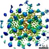
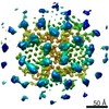
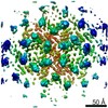
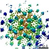
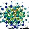
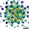
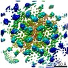
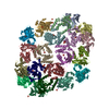
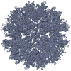
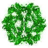
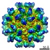
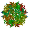
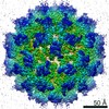
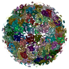
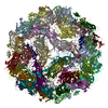
| Title | Structure of the mature RSV CA lattice: T=1 CA icosahedron | |||||||||||||||
 Components Components | Capsid protein p27, alternate cleaved 1 | |||||||||||||||
 Keywords Keywords | VIRAL PROTEIN / Retrovirus / Rous sarcoma virus / capsid protein / IP6 | |||||||||||||||
| Function / homology |  Function and homology information Function and homology informationhost cell nucleoplasm / viral procapsid maturation / host cell nucleolus / Hydrolases; Acting on peptide bonds (peptidases); Aspartic endopeptidases / viral capsid / structural constituent of virion / nucleic acid binding / aspartic-type endopeptidase activity / host cell plasma membrane / proteolysis ...host cell nucleoplasm / viral procapsid maturation / host cell nucleolus / Hydrolases; Acting on peptide bonds (peptidases); Aspartic endopeptidases / viral capsid / structural constituent of virion / nucleic acid binding / aspartic-type endopeptidase activity / host cell plasma membrane / proteolysis / zinc ion binding / membraneSimilarity search - Function | |||||||||||||||
| Biological species |  Rous sarcoma virus Rous sarcoma virus | |||||||||||||||
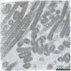
| Method | ELECTRON MICROSCOPY / single particle reconstruction / cryo EM / Resolution: 3.1 Å | |||||||||||||||
 Authors Authors | Obr, M. / Ricana, C.L. / Nikulin, N. / Feathers, J.-P.R. / Klanschnig, M. / Thader, A. / Johnson, M.C. / Vogt, V.M. / Schur, F.K.M. / Dick, R.A. | |||||||||||||||
| Funding support |  Austria, Austria,  United States, 4items United States, 4items
| |||||||||||||||
 Citation Citation | Journal: Nat Commun / Year: 2021 Title: Structure of the mature Rous sarcoma virus lattice reveals a role for IP6 in the formation of the capsid hexamer. Authors: Martin Obr / Clifton L Ricana / Nadia Nikulin / Jon-Philip R Feathers / Marco Klanschnig / Andreas Thader / Marc C Johnson / Volker M Vogt / Florian K M Schur / Robert A Dick / Abstract: Inositol hexakisphosphate (IP6) is an assembly cofactor for HIV-1. We report here that IP6 is also used for assembly of Rous sarcoma virus (RSV), a retrovirus from a different genus. IP6 is ~100-fold ...Inositol hexakisphosphate (IP6) is an assembly cofactor for HIV-1. We report here that IP6 is also used for assembly of Rous sarcoma virus (RSV), a retrovirus from a different genus. IP6 is ~100-fold more potent at promoting RSV mature capsid protein (CA) assembly than observed for HIV-1 and removal of IP6 in cells reduces infectivity by 100-fold. Here, visualized by cryo-electron tomography and subtomogram averaging, mature capsid-like particles show an IP6-like density in the CA hexamer, coordinated by rings of six lysines and six arginines. Phosphate and IP6 have opposing effects on CA in vitro assembly, inducing formation of T = 1 icosahedrons and tubes, respectively, implying that phosphate promotes pentamer and IP6 hexamer formation. Subtomogram averaging and classification optimized for analysis of pleomorphic retrovirus particles reveal that the heterogeneity of mature RSV CA polyhedrons results from an unexpected, intrinsic CA hexamer flexibility. In contrast, the CA pentamer forms rigid units organizing the local architecture. These different features of hexamers and pentamers determine the structural mechanism to form CA polyhedrons of variable shape in mature RSV particles. | |||||||||||||||
| History |
|
- Structure visualization
| Movie |
Movie viewer |
|---|---|
| Structure viewer | Molecule: MolmilJmol/JSmol |
- Downloads & links
-Download
| PDBx/mmCIF format | 7no0.cif.gz | 63.1 KB | Display | PDBx/mmCIF format |
|---|---|---|---|---|
| PDB format | pdb7no0.ent.gz | 45.9 KB | Display | PDB format |
| PDBx/mmJSON format | 7no0.json.gz | Tree view | PDBx/mmJSON format | |
| Others |  Other downloads Other downloads |
-Validation report
| Arichive directory | https://data.pdbj.org/pub/pdb/validation_reports/no/7no0ftp://data.pdbj.org/pub/pdb/validation_reports/no/7no0 | HTTPS FTP |
|---|
-Related structure data
| Related structure data | 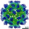 12485MC 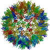 7no1C 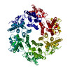 7no2C 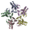 7no3C 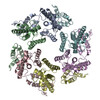 7no4C 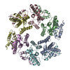 7no5C  7no6C 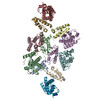 7no7C 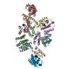 7no8C 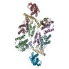 7no9C 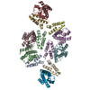 7noaC 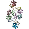 7nobC 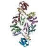 7nocC  7nodC  7noeC  7nofC  7nogC 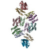 7nohC 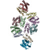 7noiC 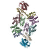 7nojC 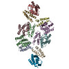 7nokC 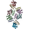 7nolC 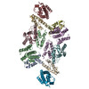 7nomC 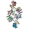 7nonC 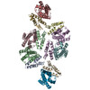 7nooC 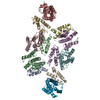 7nopC 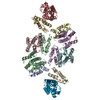 7noqC M: map data used to model this data C: citing same article ( |
|---|---|
| Similar structure data |
-Links
 PDBj
PDBj- Assembly
| Deposited unit | 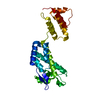
|
|---|---|
| 1 | x 60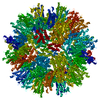
|
-Components
| #1: Protein | Mass: 24773.594 Da / Num. of mol.: 1 Source method: isolated from a genetically manipulated source Source: (gene. exp.) Rous sarcoma virus (strain Prague C) / Strain: Prague C / Gene: gag / Production host:  Escherichia coli BL21(DE3) (bacteria) / References: UniProt: P03322 Escherichia coli BL21(DE3) (bacteria) / References: UniProt: P03322 |
|---|
-Experimental details
-Experiment
| Experiment | Method: ELECTRON MICROSCOPY |
|---|---|
| EM experiment | Aggregation state: PARTICLE / 3D reconstruction method: single particle reconstruction |
- Sample preparation
| Component | Name: Rous sarcoma virus - Prague C / Type: VIRUS / Entity ID: all / Source: RECOMBINANT |
|---|---|
| Molecular weight | Experimental value: NO |
| Source (natural) | Organism: Rous sarcoma virus - Prague C |
| Source (recombinant) | Organism: Escherichia coli BL21(DE3) (bacteria) |
| Details of virus | Empty: YES / Enveloped: NO / Isolate: OTHER / Type: VIRUS-LIKE PARTICLE |
| Virus shell | Name: T=1 icosahedron |
| Buffer solution | pH: 8 |
| Buffer component | Conc.: 1 M / Name: sodium phosphate / Formula: NaPi |
| Specimen | Embedding applied: NO / Shadowing applied: NO / Staining applied: NO / Vitrification applied: YES |
| Specimen support | Grid material: COPPER / Grid mesh size: 300 divisions/in. / Grid type: C-flat-2/2 |
| Vitrification | Instrument: FEI VITROBOT MARK IV / Cryogen name: ETHANE / Humidity: 95 % / Chamber temperature: 277 K / Details: blot time= 2.5 s blot force= 0 |
- Electron microscopy imaging
| Experimental equipment |  Model: Talos Arctica / Image courtesy: FEI Company |
|---|---|
| Microscopy | Model: FEI TECNAI ARCTICA |
| Electron gun | Electron source: FIELD EMISSION GUN / Accelerating voltage: 200 kV / Illumination mode: FLOOD BEAM |
| Electron lens | Mode: BRIGHT FIELDBright-field microscopy / Nominal magnification: 63000 X / Nominal defocus max: 1500 nm / Nominal defocus min: 800 nm / Cs: 2.7 mm / C2 aperture diameter: 50 µm |
| Specimen holder | Cryogen: NITROGEN / Specimen holder model: FEI TITAN KRIOS AUTOGRID HOLDER |
| Image recording | Average exposure time: 3 sec. / Electron dose: 50 e/Å2 / Film or detector model: GATAN K3 BIOQUANTUM (6k x 4k) |
| EM imaging optics | Energyfilter name: GIF Bioquantum / Energyfilter slit width: 20 eV |
| Image scans | Width: 5760 / Height: 4092 |
- Processing
| Software | Name: PHENIX / Version: 1.17.1_3660: / Classification: refinement | ||||||||||||||||||||||||||||||||||||
|---|---|---|---|---|---|---|---|---|---|---|---|---|---|---|---|---|---|---|---|---|---|---|---|---|---|---|---|---|---|---|---|---|---|---|---|---|---|
| EM software |
| ||||||||||||||||||||||||||||||||||||
| CTF correction | Details: CTF estimation and correction was performed using GCTF in the RELION wrapper Type: PHASE FLIPPING AND AMPLITUDE CORRECTION | ||||||||||||||||||||||||||||||||||||
| Particle selection | Num. of particles selected: 150599 Details: 2394 micrographs were taken, from which 374 particles were manually picked and 2D classified to generate templates for auto-picking. Two rounds of auto-picking and 2D classification resulted ...Details: 2394 micrographs were taken, from which 374 particles were manually picked and 2D classified to generate templates for auto-picking. Two rounds of auto-picking and 2D classification resulted in 150599 extracted particles. | ||||||||||||||||||||||||||||||||||||
| Symmetry | Point symmetry: I (icosahedral) | ||||||||||||||||||||||||||||||||||||
| 3D reconstruction | Resolution: 3.1 Å / Resolution method: FSC 0.143 CUT-OFF / Num. of particles: 20498 / Symmetry type: POINT | ||||||||||||||||||||||||||||||||||||
| Atomic model building | Protocol: AB INITIO MODEL / Space: REAL Details: The CANTD and CACTD of one CA monomer of pdb 3TIR were independently placed into the EM-density using the rigid body fitting option in UCSF Chimera. Subsequently the linker connecting the ...Details: The CANTD and CACTD of one CA monomer of pdb 3TIR were independently placed into the EM-density using the rigid body fitting option in UCSF Chimera. Subsequently the linker connecting the two CA domains was joined in Coot. To account for the different monomer-monomer interactions in the RSV CA pentamer, the monomers were replicated according to the inherent 5-fold symmetry of the map and an additional ring of CACTDs was rigid-body fitted into the EM-densities of the surrounding CA pentamers. A map segment (defined by a mask extending 3 Angstrom around the rigid body fitted model) was extracted, and real-space coordinate refinement against the EM-density was performed using Phenix. This was iterated with manual model building in Coot, similar as described previously. In brief, secondary structure restraints and non-crystallographic symmetry (NCS) restraints were applied throughout all refinements. Each Phenix iteration consisted of 5 macro cycles, in which simulated annealing was performed in every macro cycle. Atomic displacement parameter (ADP) refinement was per formed at the end of each iteration. | ||||||||||||||||||||||||||||||||||||
| Atomic model building | PDB-ID: 3TIR | ||||||||||||||||||||||||||||||||||||
| Refine LS restraints |
|