Movie
Movie Controller
 Structure viewers
 About Yorodumi Papers


+Search query
-Structure paper
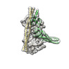
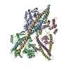
| Title | Troponin Structural Dynamics in the Native Cardiac Thin Filament Revealed by Cryo Electron Microscopy. |
|---|---|
| Journal, issue, pages | J Mol Biol, Vol. 436, Issue 6, Page 168498, Year 2024 |
| Publish date | Feb 20, 2024 |
 Authors Authors | Cristina M Risi / Betty Belknap / Jennifer Atherton / Isabella Leite Coscarella / Howard D White / P Bryant Chase / Jose R Pinto / Vitold E Galkin /  |
| PubMed Abstract | Cardiac muscle contraction occurs due to repetitive interactions between myosin thick and actin thin filaments (TF) regulated by Ca levels, active cross-bridges, and cardiac myosin-binding protein C ...Cardiac muscle contraction occurs due to repetitive interactions between myosin thick and actin thin filaments (TF) regulated by Ca levels, active cross-bridges, and cardiac myosin-binding protein C (cMyBP-C). The cardiac TF (cTF) has two nonequivalent strands, each comprised of actin, tropomyosin (Tm), and troponin (Tn). Tn shifts Tm away from myosin-binding sites on actin at elevated Ca levels to allow formation of force-producing actomyosin cross-bridges. The Tn complex is comprised of three distinct polypeptides - Ca-binding TnC, inhibitory TnI, and Tm-binding TnT. The molecular mechanism of their collective action is unresolved due to lack of comprehensive structural information on Tn region of cTF. C1 domain of cMyBP-C activates cTF in the absence of Ca to the same extent as rigor myosin. Here we used cryo-EM of native cTFs to show that cTF Tn core adopts multiple structural conformations at high and low Ca levels and that the two strands are structurally distinct. At high Ca levels, cTF is not entirely activated by Ca but exists in either partially or fully activated state. Complete dissociation of TnI C-terminus is required for full activation. In presence of cMyBP-C C1 domain, Tn core adopts a fully activated conformation, even in absence of Ca. Our data provide a structural description for the requirement of myosin to fully activate cTFs and explain increased affinity of TnC to Ca in presence of active cross-bridges. We suggest that allosteric coupling between Tn subunits and Tm is required to control actomyosin interactions. |
 External links External links | J Mol Biol / PubMed:38387550 / PubMed Central |
| Methods | EM (single particle) |
| Resolution | 5.1 - 7.1 Å |
| Structure data | EMDB-42681, PDB-8uww: EMDB-42682, PDB-8uwx: EMDB-42683, PDB-8uwy: EMDB-42800, PDB-8uyd: EMDB-42833, PDB-8uz5: EMDB-42835, PDB-8uz6: EMDB-42846, PDB-8uzx: EMDB-42847, PDB-8uzy: EMDB-42849, PDB-8v01: EMDB-42856, PDB-8v0i: EMDB-42858, PDB-8v0k: EMDB-42874, PDB-8v0y: |
| Chemicals |  ChemComp-ADP:  ChemComp-MG:  ChemComp-CA: |
| Source |
|
 Keywords Keywords |  MOTOR PROTEIN / thin filament / troponin / tropomyosin / cryo-EM / muscle structure / myosin binding protein C MOTOR PROTEIN / thin filament / troponin / tropomyosin / cryo-EM / muscle structure / myosin binding protein C |