Movie
Movie Controller

[English] 日本語
 Yorodumi
- PDB-8v01: The structure of the native cardiac thin filament troponin core i... -
+ Open data

- Basic information
| Entry | Database: PDB / ID: 8v01 | |||||||||
|---|---|---|---|---|---|---|---|---|---|---|
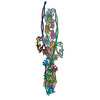
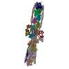
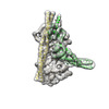
| Title | The structure of the native cardiac thin filament troponin core in Ca2+-bound fully activated state 1 from the lower strand | |||||||||
 Components Components |
| |||||||||
 Keywords Keywords |  MOTOR PROTEIN / thin filament / troponin / tropomyosin / cryo-EM / muscle structure MOTOR PROTEIN / thin filament / troponin / tropomyosin / cryo-EM / muscle structure | |||||||||
| Function / homology |  Function and homology information Function and homology informationRHOB GTPase cycle / Striated Muscle Contraction / RHOA GTPase cycle / regulation of muscle filament sliding speed / troponin T binding / actin-myosin filament sliding / cardiac Troponin complex / troponin complex / regulation of muscle contraction / transition between fast and slow fiber ...RHOB GTPase cycle / Striated Muscle Contraction / RHOA GTPase cycle / regulation of muscle filament sliding speed / troponin T binding / actin-myosin filament sliding / cardiac Troponin complex / troponin complex / regulation of muscle contraction / transition between fast and slow fiber / ventricular cardiac muscle tissue morphogenesis / myosin binding / heart contraction / mesenchyme migration / troponin I binding / skeletal muscle contraction / cardiac muscle contraction / sarcomere / filopodium / actin filament organization / actin filament / calcium-dependent protein binding / actin filament binding / actin cytoskeleton / lamellipodium / cell body / actin binding / protein heterodimerization activity / calcium ion binding / positive regulation of gene expression / protein homodimerization activity / identical protein binding / cytoplasmSimilarity search - Function | |||||||||
| Biological species |  Sus scrofa (pig) Sus scrofa (pig) | |||||||||
| Method | ELECTRON MICROSCOPY / single particle reconstruction / cryo EM / Resolution: 5.5 Å | |||||||||
 Authors Authors | Galkin, V.E. / Risi, C.M. | |||||||||
| Funding support |  United States, 2items United States, 2items
| |||||||||
 Citation Citation | Journal: J Mol Biol / Year: 2024 Title: Troponin Structural Dynamics in the Native Cardiac Thin Filament Revealed by Cryo Electron Microscopy. Authors: Cristina M Risi / Betty Belknap / Jennifer Atherton / Isabella Leite Coscarella / Howard D White / P Bryant Chase / Jose R Pinto / Vitold E Galkin / Abstract: Cardiac muscle contraction occurs due to repetitive interactions between myosin thick and actin thin filaments (TF) regulated by Ca levels, active cross-bridges, and cardiac myosin-binding protein C ...Cardiac muscle contraction occurs due to repetitive interactions between myosin thick and actin thin filaments (TF) regulated by Ca levels, active cross-bridges, and cardiac myosin-binding protein C (cMyBP-C). The cardiac TF (cTF) has two nonequivalent strands, each comprised of actin, tropomyosin (Tm), and troponin (Tn). Tn shifts Tm away from myosin-binding sites on actin at elevated Ca levels to allow formation of force-producing actomyosin cross-bridges. The Tn complex is comprised of three distinct polypeptides - Ca-binding TnC, inhibitory TnI, and Tm-binding TnT. The molecular mechanism of their collective action is unresolved due to lack of comprehensive structural information on Tn region of cTF. C1 domain of cMyBP-C activates cTF in the absence of Ca to the same extent as rigor myosin. Here we used cryo-EM of native cTFs to show that cTF Tn core adopts multiple structural conformations at high and low Ca levels and that the two strands are structurally distinct. At high Ca levels, cTF is not entirely activated by Ca but exists in either partially or fully activated state. Complete dissociation of TnI C-terminus is required for full activation. In presence of cMyBP-C C1 domain, Tn core adopts a fully activated conformation, even in absence of Ca. Our data provide a structural description for the requirement of myosin to fully activate cTFs and explain increased affinity of TnC to Ca in presence of active cross-bridges. We suggest that allosteric coupling between Tn subunits and Tm is required to control actomyosin interactions. | |||||||||
| History |
|
- Structure visualization
| Structure viewer | Molecule: MolmilJmol/JSmol |
|---|
- Downloads & links
-Download
| PDBx/mmCIF format | 8v01.cif.gz | 242.6 KB | Display | PDBx/mmCIF format |
|---|---|---|---|---|
| PDB format | pdb8v01.ent.gz | 186 KB | Display | PDB format |
| PDBx/mmJSON format | 8v01.json.gz | Tree view | PDBx/mmJSON format | |
| Others |  Other downloads Other downloads |
-Validation report
| Arichive directory | https://data.pdbj.org/pub/pdb/validation_reports/v0/8v01ftp://data.pdbj.org/pub/pdb/validation_reports/v0/8v01 | HTTPS FTP |
|---|
-Related structure data
| Related structure data |  42849MC  8uwwC  8uwxC 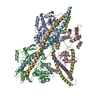 8uwyC  8uydC  8uz5C  8uz6C  8uzxC  8uzyC  8v0iC  8v0kC  8v0yC M: map data used to model this data C: citing same article ( |
|---|---|
| Similar structure data |


-Links
 PDBj
PDBj


- Assembly
| Deposited unit | 
|
|---|---|
| 1 |
|
-Components
-Protein , 5 types, 7 molecules ABCDEFG
| #1: Protein | / Cardiac muscle alpha actin 1 Mass: 42064.891 Da / Num. of mol.: 2 / Source method: isolated from a natural source / Source: (natural) Sus scrofa (pig) / References: UniProt: B6VNT8#2: Protein | | / TN-CMass: 18433.508 Da / Num. of mol.: 1 / Source method: isolated from a natural source / Source: (natural) Sus scrofa (pig) / References: UniProt: P63317#3: Protein | | / Cardiac troponin IMass: 24092.680 Da / Num. of mol.: 1 / Source method: isolated from a natural source / Source: (natural) Sus scrofa (pig) / References: UniProt: A0A4X1V710#4: Protein | | Mass: 33941.738 Da / Num. of mol.: 1 / Source method: isolated from a natural source / Source: (natural) Sus scrofa (pig) / References: UniProt: A0A5G2Q8N0#5: Protein | Mass: 32762.656 Da / Num. of mol.: 2 / Source method: isolated from a natural source / Source: (natural) Sus scrofa (pig) / References: UniProt: P42639 |
|---|
-Non-polymers , 3 types, 7 molecules 


| #6: Chemical | Adenosine diphosphate Mass: 427.201 Da / Num. of mol.: 2 / Source method: obtained synthetically / Formula: C10H15N5O10P2 / Comment: ADP, energy-carrying molecule*YM Mass: 427.201 Da / Num. of mol.: 2 / Source method: obtained synthetically / Formula: C10H15N5O10P2 / Comment: ADP, energy-carrying molecule*YM#7: Chemical |  Mass: 24.305 Da / Num. of mol.: 2 / Source method: obtained synthetically / Formula: Mg Mass: 24.305 Da / Num. of mol.: 2 / Source method: obtained synthetically / Formula: Mg#8: Chemical |  Mass: 40.078 Da / Num. of mol.: 3 / Source method: obtained synthetically / Formula: Ca Mass: 40.078 Da / Num. of mol.: 3 / Source method: obtained synthetically / Formula: Ca |
|---|
-Details
| Has ligand of interest | N |
|---|
-Experimental details
-Experiment
| Experiment | Method: ELECTRON MICROSCOPY |
|---|---|
| EM experiment | Aggregation state: FILAMENT / 3D reconstruction method: single particle reconstruction |
- Sample preparation
| Component | Name: thin filament troponin core complex / Type: COMPLEX / Entity ID: #1-#5 / Source: NATURAL |
|---|---|
| Molecular weight | Experimental value: NO |
| Source (natural) | Organism: Sus scrofa (pig) |
| Buffer solution | pH: 7.2 |
| Specimen | Embedding applied: NO / Shadowing applied: NO / Staining applied: NO / Vitrification applied: YES / Details: non-helical filament |
| Specimen support | Grid material: COPPER / Grid mesh size: 300 divisions/in. / Grid type: EMS Lacey Carbon |
| Vitrification | Instrument: FEI VITROBOT MARK IV / Cryogen name: ETHANE / Humidity: 100 % / Chamber temperature: 278 K |
- Electron microscopy imaging
| Experimental equipment |  Model: Titan Krios / Image courtesy: FEI Company |
|---|---|
| Microscopy | Model: FEI TITAN KRIOS |
| Electron gun | Electron source: FIELD EMISSION GUN / Accelerating voltage: 300 kV / Illumination mode: FLOOD BEAM |
| Electron lens | Mode: BRIGHT FIELDBright-field microscopy / Nominal defocus max: 3500 nm / Nominal defocus min: 500 nm |
| Specimen holder | Cryogen: NITROGEN / Specimen holder model: FEI TITAN KRIOS AUTOGRID HOLDER |
| Image recording | Electron dose: 34 e/Å2 / Film or detector model: GATAN K3 (6k x 4k) / Num. of real images: 24179 |
- Processing
| EM software |
| ||||||||||||||||||||||||||||||||||||||||
|---|---|---|---|---|---|---|---|---|---|---|---|---|---|---|---|---|---|---|---|---|---|---|---|---|---|---|---|---|---|---|---|---|---|---|---|---|---|---|---|---|---|
| CTF correction | Type: PHASE FLIPPING AND AMPLITUDE CORRECTION | ||||||||||||||||||||||||||||||||||||||||
| Particle selection | Num. of particles selected: 8888787 | ||||||||||||||||||||||||||||||||||||||||
| 3D reconstruction | Resolution: 5.5 Å / Resolution method: FSC 0.143 CUT-OFF / Num. of particles: 58709 / Details: Filtered to 7A / Symmetry type: POINT | ||||||||||||||||||||||||||||||||||||||||
| Atomic model building | Protocol: FLEXIBLE FIT / Space: REAL | ||||||||||||||||||||||||||||||||||||||||
| Atomic model building |
|