Movie
Movie Controller
 Structure viewers
 About Yorodumi Papers


+Search query
-Structure paper
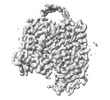
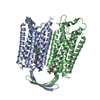
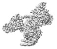
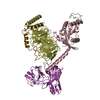
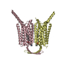
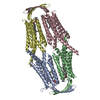
| Title | Constitutive activation mechanism of a class C GPCR. |
|---|---|
| Journal, issue, pages | Nat Struct Mol Biol, Vol. 31, Issue 4, Page 678-687, Year 2024 |
| Publish date | Feb 8, 2024 |
 Authors Authors | Jinwoo Shin / Junhyeon Park / Jieun Jeong / Jordy Homing Lam / Xingyu Qiu / Di Wu / Kuglae Kim / Joo-Youn Lee / Carol V Robinson / Jaekyung Hyun / Vsevolod Katritch / Kwang Pyo Kim / Yunje Cho /    |
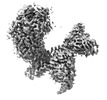
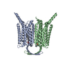
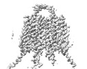
| PubMed Abstract | Class C G-protein-coupled receptors (GPCRs) are activated through binding of agonists to the large extracellular domain (ECD) followed by rearrangement of the transmembrane domains (TMDs). GPR156, a ...Class C G-protein-coupled receptors (GPCRs) are activated through binding of agonists to the large extracellular domain (ECD) followed by rearrangement of the transmembrane domains (TMDs). GPR156, a class C orphan GPCR, is unique because it lacks an ECD and exhibits constitutive activity. Impaired GPR156-G signaling contributes to loss of hearing. Here we present the cryo-electron microscopy structures of human GPR156 in the G-free and G-coupled states. We found that an endogenous phospholipid molecule is located within each TMD of the GPR156 dimer. Asymmetric binding of Gα to the phospholipid-bound GPR156 dimer restructures the first and second intracellular loops and the carboxy-terminal part of the elongated transmembrane 7 (TM7) without altering dimer conformation. Our findings reveal that GPR156 is a transducer for phospholipid signaling. Constant binding of abundant phospholipid molecules and the G-protein-induced reshaping of the cytoplasmic face provide a basis for the constitutive activation of GPR156. |
 External links External links | Nat Struct Mol Biol / PubMed:38332368 |
| Methods | EM (single particle) |
| Resolution | 2.61 - 3.33 Å |
| Structure data | EMDB-35377, PDB-8ieb: EMDB-35378, PDB-8iec: EMDB-35380, PDB-8ied: EMDB-35382, PDB-8iei: EMDB-35389, PDB-8iep: EMDB-35390, PDB-8ieq: |
| Chemicals | 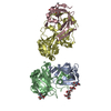 PDB-1lya: |
| Source |
|
 Keywords Keywords |  SIGNALING PROTEIN / Membrane protein / G-protein coupled receptor / Signal transduction / Phospholipid SIGNALING PROTEIN / Membrane protein / G-protein coupled receptor / Signal transduction / Phospholipid |