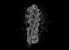
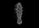
Movie
Movie Controller
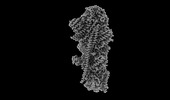
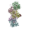
 Structure viewers
 About Yorodumi Papers


+Search query
-Structure paper
| Title | Structural basis of membrane skeleton organization in red blood cells. |
|---|---|
| Journal, issue, pages | Cell, Vol. 186, Issue 9, Page 1912-1929.e18, Year 2023 |
| Publish date | Apr 27, 2023 |
 Authors Authors | Ningning Li / Siyi Chen / Kui Xu / Meng-Ting He / Meng-Qiu Dong / Qiangfeng Cliff Zhang / Ning Gao /  |
| PubMed Abstract | The spectrin-based membrane skeleton is a ubiquitous membrane-associated two-dimensional cytoskeleton underneath the lipid membrane of metazoan cells. Mutations of skeleton proteins impair the ...The spectrin-based membrane skeleton is a ubiquitous membrane-associated two-dimensional cytoskeleton underneath the lipid membrane of metazoan cells. Mutations of skeleton proteins impair the mechanical strength and functions of the membrane, leading to several different types of human diseases. Here, we report the cryo-EM structures of the native spectrin-actin junctional complex (from porcine erythrocytes), which is a specialized short F-actin acting as the central organizational unit of the membrane skeleton. While an α-/β-adducin hetero-tetramer binds to the barbed end of F-actin as a flexible cap, tropomodulin and SH3BGRL2 together create an absolute cap at the pointed end. The junctional complex is strengthened by ring-like structures of dematin in the middle actin layers and by patterned periodic interactions with tropomyosin over its entire length. This work serves as a structural framework for understanding the assembly and dynamics of membrane skeleton and offers insights into mechanisms of various ubiquitous F-actin-binding factors in other F-actin systems. |
 External links External links | Cell / PubMed:37044097 |
| Methods | EM (single particle) |
| Resolution | 2.96 - 9.5 Å |
| Structure data | EMDB-35301, PDB-8iah: EMDB-35302, PDB-8iai: 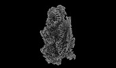 EMDB-35303: Structure of mammalian spectrin-actin junctional complex of membrane skeleton, Pointed-end segment 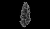 EMDB-35305: Structure of mammalian spectrin-actin junctional complex of membrane skeleton, Central segment  EMDB-35307: Structure of mammalian spectrin-actin junctional complex of membrane skeleton, Pointed-end segment, TMCC1 optimized  EMDB-35308: Structure of mammalian spectrin-actin junctional complex of membrane skeleton, Pointed-end segment, TMCC2 optimized 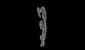 EMDB-35311: Structure of mammalian spectrin-actin junctional complex of membrane skeleton, Central segment, TMCC1 optimized  EMDB-35312: Structure of mammalian spectrin-actin junctional complex of membrane skeleton, Central segment, TMCC2 optimized 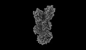 EMDB-35317: Structure of mammalian spectrin-actin junctional complex of membrane skeleton, State I, Barbed-end segment  EMDB-35318: Structure of mammalian spectrin-actin junctional complex of membrane skeleton, State I, Barbed-end segment, adducin optimized 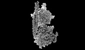 EMDB-35319: Structure of mammalian spectrin-actin junctional complex of membrane skeleton, State I, Barbed-end segment, TMCC1 optimized  EMDB-35320: Structure of mammalian spectrin-actin junctional complex of membrane skeleton, State I, Barbed-end segment, TMCC2 optimized  EMDB-35321: Structure of mammalian spectrin-actin junctional complex of membrane skeleton, State I, Barbed-end segment, the second spectrin repeat dimer optimized 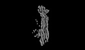 EMDB-35322: Structure of mammalian spectrin-actin junctional complex of membrane skeleton, State I, Barbed-end segment, the first two spectrin repeat dimers optimized 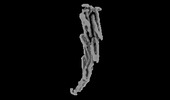 EMDB-35324: Structure of mammalian spectrin-actin junctional complex of membrane skeleton, State I, Barbed-end segment, the third to fifth spectrin repeat dimers optimized  EMDB-35325: Structure of mammalian spectrin-actin junctional complex of membrane skeleton, State II, Barbed-end segment 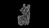 EMDB-35327: Structure of mammalian spectrin-actin junctional complex of membrane skeleton, State II, Barbed-end segment, adducin/TMCC1 optimized EMDB-35329, PDB-8ib2: |
| Chemicals |  ChemComp-ADP: |
| Source |
|
 Keywords Keywords |  MEMBRANE PROTEIN / Macrocomplex / membrane skeleton / spectrin-actin junction MEMBRANE PROTEIN / Macrocomplex / membrane skeleton / spectrin-actin junction |