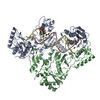
 ムービー
ムービー コントローラー
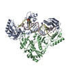
 構造ビューア
 EMN検索について


-検索条件
-検索結果
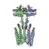
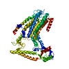
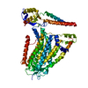
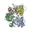
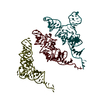
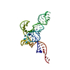
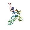
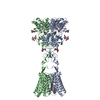
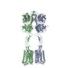
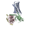
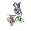
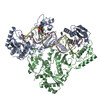
検索 (著者・登録者: fu, & j.)の結果70件中、1から50件目までを表示しています
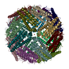
PDB-8wcn: 
Cryo-EM structure of PAO1-ImcA with GMPCPP

PDB-8ff2:
Amyloid-beta (1-40) fibrils derived from a CAA patient

PDB-8ff3:
Amyloid-beta (1-40) fibrils derived from familial Dutch-type CAA patient (population B)

PDB-8jd9:
Cyro-EM structure of the Na+/H+ antipoter SOS1 from Arabidopsis thaliana,class1

PDB-8jda:
Cyro-EM structure of the Na+/H+ antipoter SOS1 from Arabidopsis thaliana,class2
PDB-8ehw:
cryo-EM structure of TMEM63A in nanodisc
PDB-8ehx:
cryo-EM structure of TMEM63B in LMNG
PDB-7ypo:
Cryo-EM structure of baculovirus LEF-3 in complex with ssDNA

PDB-7ypq:
Cryo-EM structure of one baculovirus LEF-3 molecule in complex with ssDNA
PDB-7t9p:
Cryo-EM structure of Human Enterovirus D68 US/MO/14-18947 strain native virion
PDB-7taf:
Cryo-EM structure of Human Enterovirus D68 US/MO/14-18947 strain virion in complex with inhibitor 11526092

PDB-7tag:
Cryo-EM structure of Human Enterovirus D68 US/MO/14-18947 strain virion in complex with pleconaril

PDB-7tah:
Cryo-EM structure of Human Enterovirus D68 US/MO/14-18947 strain in complex with inhibitor 11526091 (no/low occupancy-no inhibitor modeled)

PDB-7taj:
Cryo-EM structure of Human Enterovirus D68 US/MO/14-18947 strain in complex with inhibitor 11526093 (no/low occupancy-no inhibitor modeled)
PDB-8ehg:
Rabbit muscle aldolase determined using single-particle cryo-EM with Apollo camera.
PDB-8emq:
Mouse apoferritin heavy chain with zinc determined using single-particle cryo-EM with Apollo camera.
PDB-8en7:
Mouse apoferritin heavy chain without zinc determined using single-particle cryo-EM with Apollo camera.
PDB-7xn4:
Cryo-EM structure of CopC-CaM-caspase-3 with NAD+
PDB-7xn5:
Cryo-EM structure of CopC-CaM-caspase-3 with ADPR

PDB-7xn6:
Cryo-EM structure of CopC-CaM-caspase-3 with ADPR-deacylization

PDB-8h0x:
Structure of SARS-CoV-1 Spike Protein with Engineered x1 Disulfide (S370C and D967C), Locked-1 Conformation

PDB-8h0y:
Structure of SARS-CoV-1 Spike Protein with Engineered x1 Disulfide (S370C and D967C), Locked-112 Conformation

PDB-8h0z:
Structure of SARS-CoV-1 Spike Protein with Engineered x1 Disulfide (S370C and D967C), Locked-122 Conformation
PDB-8h11:
Structure of SARS-CoV-1 Spike Protein with Engineered x1 Disulfide (S370C and D967C), Closed Conformation

PDB-8h12:
Structure of SARS-CoV-1 Spike Protein with Engineered x2 Disulfide (G400C and V969C), Locked-2 Conformation
PDB-8h15:
Structure of SARS-CoV-1 Spike Protein (S/native) at pH 5.5, Closed Conformation
PDB-8h16:
Structure of SARS-CoV-1 Spike Protein (S/native) at pH 5.5, Open Conformation
PDB-8h10:
Structure of SARS-CoV-1 Spike Protein with Engineered x1 Disulfide (S370C and D967C), Locked-2 Conformation
PDB-8h13:
Structure of SARS-CoV-1 Spike Protein with Engineered x2 Disulfide (G400C and V969C), Closed Conformation

PDB-8h14:
Structure of SARS-CoV-1 Spike Protein with Engineered x3 Disulfide (D414C and V969C), Locked-1 Conformation
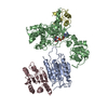
PDB-7ur5:
allo-tRNAUTu1 in the A, P, and E sites of the E. coli ribosome
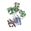
PDB-7uri:
allo-tRNAUTu1A in the A site of the E. coli ribosome
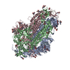
PDB-7urm:
allo-tRNAUTu1A in the P site of the E. coli ribosome

PDB-7sil:
Structure of positive allosteric modulator-bound active human calcium-sensing receptor
PDB-7sim:
Structure of positive allosteric modulator-free active human calcium-sensing receptor
PDB-7sin:
Structure of negative allosteric modulator-bound inactive human calcium-sensing receptor
PDB-7f6h:
Cryo-EM structure of human bradykinin receptor BK2R in complex Gq proteins and bradykinin
PDB-7f6i:
Cryo-EM structure of human bradykinin receptor BK2R in complex Gq proteins and kallidin
PDB-7kjv:
Structure of HIV-1 reverse transcriptase initiation complex core
PDB-7kjw:
Structure of HIV-1 reverse transcriptase initiation complex core with efavirenz
PDB-7kjx:
Structure of HIV-1 reverse transcriptase initiation complex core with nevirapine
PDB-6wds:
Enterovirus D68 in complex with human monoclonal antibody EV68-159
PDB-6wdt:
Enterovirus D68 in complex with human monoclonal antibody EV68-228
PDB-6wiv:
Structure of human GABA(B) receptor in an inactive state

PDB-6wej:
Structure of cGMP-unbound WT TAX-4 reconstituted in lipid nanodiscs

PDB-6wek:
Structure of cGMP-bound WT TAX-4 reconstituted in lipid nanodiscs

PDB-6wel:
Structure of cGMP-unbound F403V/V407A mutant TAX-4 reconstituted in lipid nanodiscs
PDB-6vxf:
Structure of apo-closed ABCG2

PDB-6osk:
RF1 accommodated 70S complex at 60 ms
PDB-6osq:
RF1 accommodated state bound Release complex 70S at long incubation time point
ページ:
 wwPDBはEMDBデータモデルのバージョン3へ移行します
