 Movie
Movie Controller
 Structure viewers
 About EMN search


-Search query
-Search result
Showing 1 - 50 of 81 items for (author: gan & sy)
EMDB-19406: 
Structure of the human DDB1-DDA1-DCAF15 E3 ubiquitin ligase bound to compound furan 12
EMDB-19407:
Structure of the human DDB1-DDA1-DCAF15 E3 ubiquitin ligase bound to compound furan 24
PDB-8rox:
Structure of the human DDB1-DDA1-DCAF15 E3 ubiquitin ligase bound to compound furan 12
PDB-8roy:
Structure of the human DDB1-DDA1-DCAF15 E3 ubiquitin ligase bound to compound furan 24
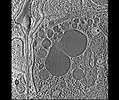
EMDB-28138:
3D reconstruction of the apical complex of Plasmodium falciparum (3D7) free merozoite
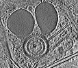
EMDB-28141:
3D reconstruction of the apical complex of Plasmodium falciparum (3D7) free merozoite
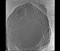
EMDB-28142:
3D Reconstruction of Plasmodium falciparum (3D7) free merozoite
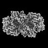
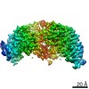
EMDB-42804:
Structure of nucleotide-free Pediculus humanus (Ph) PINK1 dimer

EMDB-42806:
Structure of AMP-PNP-bound Pediculus humanus (Ph) PINK1 dimer

EMDB-42807:
Structure of ADP-bound and phosphorylated Pediculus humanus (Ph) PINK1 dimer
PDB-8uyf:
Structure of nucleotide-free Pediculus humanus (Ph) PINK1 dimer

PDB-8uyh:
Structure of AMP-PNP-bound Pediculus humanus (Ph) PINK1 dimer

PDB-8uyi:
Structure of ADP-bound and phosphorylated Pediculus humanus (Ph) PINK1 dimer
EMDB-36887:
Structure of GPR34-Gi complex

EMDB-28125:
Plasmodium falciparum merozoites apical 2-ring units

EMDB-28126:
Toxoplasma apical rings
EMDB-28910:
Glycan-Base ConC Env Trimer

PDB-8f7t:
Glycan-Base ConC Env Trimer

EMDB-27630:
Structure of the PEAK3/14-3-3 complex

EMDB-27684:
Structure of the PEAK3 pseudokinase homodimer
PDB-8dp5:
Structure of the PEAK3/14-3-3 complex

PDB-8ds6:
Structure of the PEAK3 pseudokinase homodimer
EMDB-29396:
Antibody vFP53.02 in complex with HIV-1 envelope trimer BG505 DS-SOSIP
EMDB-29836:
vFP52.02 Fab in complex with BG505 DS-SOSIP Env trimer
EMDB-29880:
Cryo-EM structure of vFP49.02 Fab in complex with HIV-1 Env BG505 DS-SOSIP.664 (conformation 1)
EMDB-29881:
Cryo-EM structure of vFP49.02 Fab in complex with HIV-1 Env BG505 DS-SOSIP.664 (conformation 2)
EMDB-29882:
Cryo-EM structure of vFP49.02 Fab in complex with HIV-1 Env BG505 DS-SOSIP.664 (conformation 3)
EMDB-29905:
vFP48.02 Fab in complex with BG505 DS-SOSIP Env trimer

PDB-8fr6:
Antibody vFP53.02 in complex with HIV-1 envelope trimer BG505 DS-SOSIP

PDB-8g85:
vFP52.02 Fab in complex with BG505 DS-SOSIP Env trimer
PDB-8g9w:
Cryo-EM structure of vFP49.02 Fab in complex with HIV-1 Env BG505 DS-SOSIP.664 (conformation 1)
PDB-8g9x:
Cryo-EM structure of vFP49.02 Fab in complex with HIV-1 Env BG505 DS-SOSIP.664 (conformation 2)

PDB-8g9y:
Cryo-EM structure of vFP49.02 Fab in complex with HIV-1 Env BG505 DS-SOSIP.664 (conformation 3)

PDB-8gas:
vFP48.02 Fab in complex with BG505 DS-SOSIP Env trimer
EMDB-28139:
Toxoplasma gondii apical complex (ionophore stimulated)
EMDB-28140:
Toxoplasma gondii apical complex (non-stimulated)
EMDB-13776:
Structure of formaldehyde cross-linked SARS-CoV-2 S glycoprotein
PDB-7q1z:
Structure of formaldehyde cross-linked SARS-CoV-2 S glycoprotein

EMDB-25677:
Structure of dimeric phosphorylated Pediculus humanus (Ph) PINK1
EMDB-25678:
Structure of dimeric phosphorylated Pediculus humanus (Ph) PINK1 with kinked alpha-C helix in chain B

EMDB-25679:
Structure of dimeric phosphorylated Pediculus humanus (Ph) PINK1 with extended alpha-C helix in chain B

EMDB-25680:
Structure of dodecameric unphosphorylated Pediculus humanus (Ph) PINK1 D357A mutant

EMDB-25681:
Structure of dimeric unphosphorylated Pediculus humanus (Ph) PINK1 D357A mutant

PDB-7t4k:
Structure of dimeric phosphorylated Pediculus humanus (Ph) PINK1 with kinked alpha-C helix in chain B

PDB-7t4l:
Structure of dimeric phosphorylated Pediculus humanus (Ph) PINK1 with extended alpha-C helix in chain B

PDB-7t4m:
Structure of dodecameric unphosphorylated Pediculus humanus (Ph) PINK1 D357A mutant

PDB-7t4n:
Structure of dimeric unphosphorylated Pediculus humanus (Ph) PINK1 D357A mutant
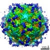
EMDB-30883:
Cryo-EM structure of Dengue virus serotype 1 strain WestPac 74 in complex with human antibody 1C19 Fab at 37 deg C (Class 1 particle)
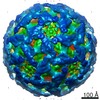
EMDB-30884:
Cryo-EM structure of Dengue virus serotype 1 strain WestPac 74 in complex with human antibody 1C19 Fab at 37 deg C (Class 2 particle)
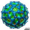
EMDB-30885:
Cryo-EM map of Dengue virus serotype 2 strain PVP94/07 in complex with human antibody 1C19 Fab at 37 deg C
Pages:
 wwPDB to switch to version 3 of the EMDB data model
