National Institutes of Health/National Institute of General Medical Sciences (NIH/NIGMS)
R01GM079429
United States
National Institutes of Health/National Institute Of Allergy and Infectious Diseases (NIH/NIAID)
DP2HL137186
United States
Citation
Journal: mBio / Year: 2024 Title: Cryogenic electron tomography reveals novel structures in the apical complex of . Authors: Stella Y Sun / Li-Av Segev-Zarko / Grigore D Pintilie / Chi Yong Kim / Sophia R Staggers / Michael F Schmid / Elizabeth S Egan / Wah Chiu / John C Boothroyd / Abstract: Intracellular infectious agents, like the malaria parasite, , face the daunting challenge of how to invade a host cell. This problem may be even harder when the host cell in question is the ...Intracellular infectious agents, like the malaria parasite, , face the daunting challenge of how to invade a host cell. This problem may be even harder when the host cell in question is the enucleated red blood cell, which lacks the host machinery co-opted by many pathogens for internalization. Evolution has provided and related single-celled parasites within the phylum Apicomplexa with a collection of organelles at their apical end that mediate invasion. This apical complex includes at least two sets of secretory organelles, micronemes and rhoptries, and several structural features like apical rings and a putative pore through which proteins may be introduced into the host cell during invasion. We perform cryogenic electron tomography (cryo-ET) equipped with Volta Phase Plate on isolated and vitrified merozoites to visualize the apical machinery. Through tomographic reconstruction of cellular compartments, we see new details of known structures like the rhoptry tip interacting directly with a rosette resembling the recently described rhoptry secretory apparatus (RSA) or with an apical vesicle docked beneath the RSA. Subtomogram averaging reveals that the apical rings have a fixed number of repeating units, each of which is similar in overall size and shape to the units in the apical rings of tachyzoites of . Comparison of these polar rings in and parasites also reveals them to have a structurally conserved assembly pattern. These results provide new insight into the essential and structurally conserved features of this remarkable machinery used by apicomplexan parasites to invade their respective host cells. IMPORTANCE: Malaria is an infectious disease caused by parasites of the genus and is a leading cause of morbidity and mortality globally. Upon infection, parasites invade and replicate in red blood ...IMPORTANCE: Malaria is an infectious disease caused by parasites of the genus and is a leading cause of morbidity and mortality globally. Upon infection, parasites invade and replicate in red blood cells, where they are largely protected from the immune system. To enter host cells, the parasites employ a specialized apparatus at their anterior end. In this study, advanced imaging techniques like cryogenic electron tomography (cryo-ET) and Volta Phase Plate enable unprecedented visualization of whole merozoites, revealing previously unknown structural details of their invasion machinery. Key findings include new insights into the structural conservation of apical rings shared between and its apicomplexan cousin, . These discoveries shed light on the essential and conserved elements of the invasion machinery used by these pathogens. Moreover, the research provides a foundation for understanding the molecular mechanisms underlying parasite-host interactions, potentially informing strategies for combating diseases caused by apicomplexan parasites.
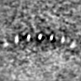
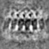
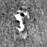
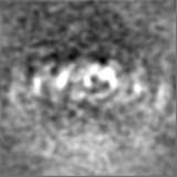
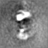
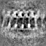
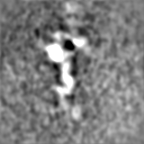
This map is obtained by positioning the reconstructed full map at multiple positions around the rings in tomograms. This map is cropped to show a small segment of the entire rings.
Film or detector model: GATAN K2 SUMMIT (4k x 4k) / Detector mode: COUNTING / Average electron dose: 1.8 e/Å2
Experimental equipment
Model: Talos Arctica / Image courtesy: FEI Company
-
Image processing
Extraction
Number tomograms: 59 / Number images used: 335
Final angle assignment
Type: NOT APPLICABLE
Final reconstruction
Applied symmetry - Point group: C1 (asymmetric) / Resolution.type: BY AUTHOR / Resolution: 59.67 Å / Resolution method: OTHER / Number subtomograms used: 335
+
About Yorodumi
-
News
-
Feb 9, 2022. New format data for meta-information of EMDB entries
New format data for meta-information of EMDB entries
Version 3 of the EMDB header file is now the official format.
The previous official version 1.9 will be removed from the archive.
In the structure databanks used in Yorodumi, some data are registered as the other names, "COVID-19 virus" and "2019-nCoV". Here are the details of the virus and the list of structure data.
Jan 31, 2019. EMDB accession codes are about to change! (news from PDBe EMDB page)
EMDB accession codes are about to change! (news from PDBe EMDB page)
The allocation of 4 digits for EMDB accession codes will soon come to an end. Whilst these codes will remain in use, new EMDB accession codes will include an additional digit and will expand incrementally as the available range of codes is exhausted. The current 4-digit format prefixed with “EMD-” (i.e. EMD-XXXX) will advance to a 5-digit format (i.e. EMD-XXXXX), and so on. It is currently estimated that the 4-digit codes will be depleted around Spring 2019, at which point the 5-digit format will come into force.
The EM Navigator/Yorodumi systems omit the EMD- prefix.
Related info.:Q: What is EMD? / ID/Accession-code notation in Yorodumi/EM Navigator
Yorodumi is a browser for structure data from EMDB, PDB, SASBDB, etc.
This page is also the successor to EM Navigator detail page, and also detail information page/front-end page for Omokage search.
The word "yorodu" (or yorozu) is an old Japanese word meaning "ten thousand". "mi" (miru) is to see.
Related info.:EMDB / PDB / SASBDB / Comparison of 3 databanks / Yorodumi Search / Aug 31, 2016. New EM Navigator & Yorodumi / Yorodumi Papers / Jmol/JSmol / Function and homology information / Changes in new EM Navigator and Yorodumi
 Movie
Movie Controller


 Open data
 Basic information
 Map data
 Sample
 Keywords
 Cryo-electron tomography / Subtomogram averaging / Apical ring /
 Authors
 United States, 2 items
  Citation
 Structure visualization
 Downloads & links
 EMDB map data format
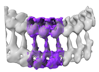 emd_28126.png
 http://ftp.pdbj.org/pub/emdb/structures/EMD-28126
 Z
Z Y
Y X
X


 Sample components
 Processing
 Electron microscopy
