 Movie
Movie Controller
 Structure viewers
 About Yorodumi Papers


+Search query
-Structure paper
| Title | Cryogenic electron tomography reveals novel structures in the apical complex of . |
|---|---|
| Journal, issue, pages | mBio, Vol. 15, Issue 4, Page e0286423, Year 2024 |
| Publish date | Apr 10, 2024 |
 Authors Authors | Stella Y Sun / Li-Av Segev-Zarko / Grigore D Pintilie / Chi Yong Kim / Sophia R Staggers / Michael F Schmid / Elizabeth S Egan / Wah Chiu / John C Boothroyd /  |
| PubMed Abstract | Intracellular infectious agents, like the malaria parasite, , face the daunting challenge of how to invade a host cell. This problem may be even harder when the host cell in question is the ...Intracellular infectious agents, like the malaria parasite, , face the daunting challenge of how to invade a host cell. This problem may be even harder when the host cell in question is the enucleated red blood cell, which lacks the host machinery co-opted by many pathogens for internalization. Evolution has provided and related single-celled parasites within the phylum Apicomplexa with a collection of organelles at their apical end that mediate invasion. This apical complex includes at least two sets of secretory organelles, micronemes and rhoptries, and several structural features like apical rings and a putative pore through which proteins may be introduced into the host cell during invasion. We perform cryogenic electron tomography (cryo-ET) equipped with Volta Phase Plate on isolated and vitrified merozoites to visualize the apical machinery. Through tomographic reconstruction of cellular compartments, we see new details of known structures like the rhoptry tip interacting directly with a rosette resembling the recently described rhoptry secretory apparatus (RSA) or with an apical vesicle docked beneath the RSA. Subtomogram averaging reveals that the apical rings have a fixed number of repeating units, each of which is similar in overall size and shape to the units in the apical rings of tachyzoites of . Comparison of these polar rings in and parasites also reveals them to have a structurally conserved assembly pattern. These results provide new insight into the essential and structurally conserved features of this remarkable machinery used by apicomplexan parasites to invade their respective host cells. IMPORTANCE: Malaria is an infectious disease caused by parasites of the genus and is a leading cause of morbidity and mortality globally. Upon infection, parasites invade and replicate in red blood cells, where they are largely protected from the immune system. To enter host cells, the parasites employ a specialized apparatus at their anterior end. In this study, advanced imaging techniques like cryogenic electron tomography (cryo-ET) and Volta Phase Plate enable unprecedented visualization of whole merozoites, revealing previously unknown structural details of their invasion machinery. Key findings include new insights into the structural conservation of apical rings shared between and its apicomplexan cousin, . These discoveries shed light on the essential and conserved elements of the invasion machinery used by these pathogens. Moreover, the research provides a foundation for understanding the molecular mechanisms underlying parasite-host interactions, potentially informing strategies for combating diseases caused by apicomplexan parasites. |
 External links External links | mBio / PubMed:38456679 / PubMed Central |
| Methods | EM (subtomogram averaging) / EM (tomography) |
| Resolution | 41.0 - 59.67 Å |
| Structure data |  EMDB-28125: Plasmodium falciparum merozoites apical 2-ring units  EMDB-28126: Toxoplasma apical rings 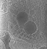 EMDB-28138: 3D reconstruction of the apical complex of Plasmodium falciparum (3D7) free merozoite 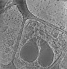 EMDB-28141: 3D reconstruction of the apical complex of Plasmodium falciparum (3D7) free merozoite 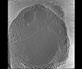 EMDB-28142: 3D Reconstruction of Plasmodium falciparum (3D7) free merozoite |
| Source |
|

 Plasmodium falciparum 3D7 (eukaryote)