 Movie
Movie Controller
 Structure viewers
 About EMN search


-Search query
-Search result
Showing 1 - 50 of 439 items for (author: white & p)
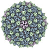
EMDB-40271: 
Mycobacterium phage Adjutor
Method: single particle / : Podgorski JM, White SJ
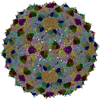
PDB-8saj:
Mycobacterium phage Adjutor
Method: single particle / : Podgorski JM, White SJ

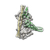
EMDB-42681:
The structure of the native cardiac thin filament troponin core in Ca2+-free state from the upper strand
Method: single particle / : Galkin VE, Risi CM

EMDB-42682:
The structure of the native cardiac thin filament troponin core in Ca2+-free tilted state from the upper strand
Method: single particle / : Galkin VE, Risi CM
EMDB-42683:
The structure of the native cardiac thin filament troponin core in Ca2+-free rotated state from the upper strand
Method: single particle / : Galkin VE, Risi CM

EMDB-42800:
The structure of the native cardiac thin filament troponin core in Ca2+-free state from the lower strand
Method: single particle / : Galkin VE, Risi CM

EMDB-42833:
The structure of the native cardiac thin filament troponin core in Ca2+-free rotated state from the lower strand
Method: single particle / : Galkin VE, Risi CM

EMDB-42835:
The structure of the native cardiac thin filament troponin core in Ca2+-free tilted state from the lower strand
Method: single particle / : Galkin VE, Risi CM

EMDB-42846:
The structure of the native cardiac thin filament troponin core in Ca2+-bound fully activated state from the upper strand
Method: single particle / : Galkin VE, Risi CM

EMDB-42847:
The structure of the native cardiac thin filament troponin core in Ca2+-bound partially activated state from the upper strand
Method: single particle / : Galkin VE, Risi CM

EMDB-42849:
The structure of the native cardiac thin filament troponin core in Ca2+-bound fully activated state 1 from the lower strand
Method: single particle / : Galkin VE, Risi CM

EMDB-42856:
The structure of the native cardiac thin filament troponin core in Ca2+-bound fully activated state 2 from the lower strand
Method: single particle / : Galkin VE, Risi CM

EMDB-42858:
The structure of the native cardiac thin filament troponin core in Ca2+-bound partially activated state from the lower strand
Method: single particle / : Galkin VE, Risi CM

EMDB-42874:
The structure of the native cardiac thin filament troponin core in Ca2+-free state from the upper strand activated by the C1-domain of cardiac myosin binding protein C
Method: single particle / : Galkin VE, Risi CM

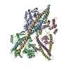
PDB-8uww:
The structure of the native cardiac thin filament troponin core in Ca2+-free state from the upper strand
Method: single particle / : Galkin VE, Risi CM

PDB-8uwx:
The structure of the native cardiac thin filament troponin core in Ca2+-free tilted state from the upper strand
Method: single particle / : Galkin VE, Risi CM
PDB-8uwy:
The structure of the native cardiac thin filament troponin core in Ca2+-free rotated state from the upper strand
Method: single particle / : Galkin VE, Risi CM

PDB-8uyd:
The structure of the native cardiac thin filament troponin core in Ca2+-free state from the lower strand
Method: single particle / : Galkin VE, Risi CM

PDB-8uz5:
The structure of the native cardiac thin filament troponin core in Ca2+-free rotated state from the lower strand
Method: single particle / : Galkin VE, Risi CM

PDB-8uz6:
The structure of the native cardiac thin filament troponin core in Ca2+-free tilted state from the lower strand
Method: single particle / : Galkin VE, Risi CM

PDB-8uzx:
The structure of the native cardiac thin filament troponin core in Ca2+-bound fully activated state from the upper strand
Method: single particle / : Galkin VE, Risi CM

PDB-8uzy:
The structure of the native cardiac thin filament troponin core in Ca2+-bound partially activated state from the upper strand
Method: single particle / : Galkin VE, Risi CM

PDB-8v01:
The structure of the native cardiac thin filament troponin core in Ca2+-bound fully activated state 1 from the lower strand
Method: single particle / : Galkin VE, Risi CM

PDB-8v0i:
The structure of the native cardiac thin filament troponin core in Ca2+-bound fully activated state 2 from the lower strand
Method: single particle / : Galkin VE, Risi CM

PDB-8v0k:
The structure of the native cardiac thin filament troponin core in Ca2+-bound partially activated state from the lower strand
Method: single particle / : Galkin VE, Risi CM

PDB-8v0y:
The structure of the native cardiac thin filament troponin core in Ca2+-free state from the upper strand activated by the C1-domain of cardiac myosin binding protein C
Method: single particle / : Galkin VE, Risi CM
EMDB-35809:
Cellular components in INS-1E cell periphery
Method: electron tomography / : Li W, Li A, Yu B, Zhang X, Liu X, White K, Stevens R, Baumeister W, Sali A, Jasnin M, Sun L
EMDB-35839:
Cellular components at INS-1E cell periphery under second phase of glucose-stimulated insulin secretion
Method: electron tomography / : Li W, Li A, Yu B, Zhang X, Liu X, White K, Stevens R, Baumeister W, Sali A, Jasnin M, Sun L
EMDB-35840:
Cellular components at INS-1E cell periphery under first phase of glucose-stimulated insulin secretion
Method: electron tomography / : Li W, Li A, Yu B, Zhang X, Liu X, White K, Stevens R, Baumeister W, Sali A, Jasnin M, Sun L
EMDB-35841:
Cellular components at INS-1E cell periphery under basal condition
Method: electron tomography / : Li W, Li A, Yu B, Zhang X, Liu X, White K, Stevens R, Baumeister W, Sali A, Jasnin M, Sun L

EMDB-35842:
Cellular components at INS-1E cell periphery under basal condition
Method: electron tomography / : Li W, Li A, Yu B, Zhang X, Liu X, White K, Stevens R, Baumeister W, Sali A, Jasnin M, Sun L

EMDB-35843:
Cellular components at INS-1E cell periphery under basal condition
Method: electron tomography / : Li W, Li A, Yu B, Zhang X, Liu X, White K, Stevens R, Baumeister W, Sali A, Jasnin M, Sun L

EMDB-35844:
Cellular components at INS-1E cell periphery under basal condition
Method: electron tomography / : Li W, Li A, Yu B, Zhang X, Liu X, White K, Stevens R, Baumeister W, Sali A, Jasnin M, Sun L
EMDB-35845:
Cellular components at INS-1E cell periphery under basal condition
Method: electron tomography / : Li W, Li A, Yu B, Zhang X, Liu X, White K, Stevens R, Baumeister W, Sali A, Jasnin M, Sun L
EMDB-35846:
Cellular components at INS-1E cell periphery under basal condition
Method: electron tomography / : Li W, Li A, Yu B, Zhang X, Liu X, White K, Stevens R, Baumeister W, Sali A, Jasnin M, Sun L
EMDB-35847:
Cellular components at INS-1E cell periphery under basal condition
Method: electron tomography / : Li W, Li A, Yu B, Zhang X, Liu X, White K, Stevens R, Baumeister W, Sali A, Jasnin M, Sun L
EMDB-35848:
Cellular components at INS-1E cell periphery under second phase of glucose-stimulated insulin secretion
Method: electron tomography / : Li W, Li A, Yu B, Zhang X, Liu X, White K, Stevens R, Baumeister W, Sali A, Jasnin M, Sun L
EMDB-35849:
Cellular components at INS-1E cell periphery under second phase of glucose-stimulated insulin secretion
Method: electron tomography / : Li W, Li A, Yu B, Zhang X, Liu X, White K, Stevens R, Baumeister W, Sali A, Jasnin M, Sun L
EMDB-35850:
Cellular components at INS-1E cell periphery under second phase of glucose-stimulated insulin secretion
Method: electron tomography / : Li W, Li A, Yu B, Zhang X, Liu X, White K, Stevens R, Baumeister W, Sali A, Jasnin M, Sun L
EMDB-35851:
Cellular components at INS-1E cell periphery under second phase of glucose-stimulated insulin secretion
Method: electron tomography / : Li W, Li A, Yu B, Zhang X, Liu X, White K, Stevens R, Baumeister W, Sali A, Jasnin M, Sun L

EMDB-35852:
Cellular components at INS-1E cell periphery under second phase of glucose-stimulated insulin secretion
Method: electron tomography / : Li W, Li A, Yu B, Zhang X, Liu X, White K, Stevens R, Baumeister W, Sali A, Jasnin M, Sun L
EMDB-35853:
Cellular components at INS-1E cell periphery under second phase of glucose-stimulated insulin secretion
Method: electron tomography / : Li W, Li A, Yu B, Zhang X, Liu X, White K, Stevens R, Baumeister W, Sali A, Jasnin M, Sun L
EMDB-35854:
Cellular components at INS-1E cell periphery under second phase of glucose-stimulated insulin secretion
Method: electron tomography / : Li W, Li A, Yu B, Zhang X, Liu X, White K, Stevens R, Baumeister W, Sali A, Jasnin M, Sun L
EMDB-35855:
Cellular components at INS-1E cell periphery under first phase of glucose-stimulated insulin secretion
Method: electron tomography / : Li W, Li A, Yu B, Zhang X, Liu X, White K, Stevens R, Baumeister W, Sali A, Jasnin M, Sun L
EMDB-35856:
Cellular components at INS-1E cell periphery under first phase of glucose-stimulated insulin secretion
Method: electron tomography / : Li W, Li A, Yu B, Zhang X, Liu X, White K, Stevens R, Baumeister W, Sali A, Jasnin M, Sun L
EMDB-35857:
Cellular components at INS-1E cell periphery under first phase of glucose-stimulated insulin secretion
Method: electron tomography / : Li W, Li A, Yu B, Zhang X, Liu X, White K, Stevens R, Baumeister W, Sali A, Jasnin M, Sun L
EMDB-35858:
Cellular components at INS-1E cell periphery under first phase of glucose-stimulated insulin secretion
Method: electron tomography / : Li W, Li A, Yu B, Zhang X, Liu X, White K, Stevens R, Baumeister W, Sali A, Jasnin M, Sun L
EMDB-35859:
Cellular components at INS-1E cell periphery under first phase of glucose-stimulated insulin secretion
Method: electron tomography / : Li W, Li A, Yu B, Zhang X, Liu X, White K, Stevens R, Baumeister W, Sali A, Jasnin M, Sun L
EMDB-35860:
Cellular components at INS-1E cell periphery under first phase of glucose-stimulated insulin secretion
Method: electron tomography / : Li W, Li A, Yu B, Zhang X, Liu X, White K, Stevens R, Baumeister W, Sali A, Jasnin M, Sun L
EMDB-35861:
Cellular components at INS-1E cell periphery under first phase of glucose-stimulated insulin secretion
Method: electron tomography / : Li W, Li A, Yu B, Zhang X, Liu X, White K, Stevens R, Baumeister W, Sali A, Jasnin M, Sun L
Pages:
 wwPDB to switch to version 3 of the EMDB data model
