 Movie
Movie Controller
 Structure viewers
 About EMN search


-Search query
-Search result
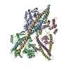
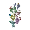
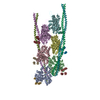
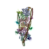
Showing all 38 items for (author: galkin, & v.e.)

PDB-8uww: 
The structure of the native cardiac thin filament troponin core in Ca2+-free state from the upper strand
Method: single particle / : Galkin VE, Risi CM

PDB-8uwx:
The structure of the native cardiac thin filament troponin core in Ca2+-free tilted state from the upper strand
Method: single particle / : Galkin VE, Risi CM
PDB-8uwy:
The structure of the native cardiac thin filament troponin core in Ca2+-free rotated state from the upper strand
Method: single particle / : Galkin VE, Risi CM

PDB-8uyd:
The structure of the native cardiac thin filament troponin core in Ca2+-free state from the lower strand
Method: single particle / : Galkin VE, Risi CM

PDB-8uz5:
The structure of the native cardiac thin filament troponin core in Ca2+-free rotated state from the lower strand
Method: single particle / : Galkin VE, Risi CM

PDB-8uz6:
The structure of the native cardiac thin filament troponin core in Ca2+-free tilted state from the lower strand
Method: single particle / : Galkin VE, Risi CM

PDB-8uzx:
The structure of the native cardiac thin filament troponin core in Ca2+-bound fully activated state from the upper strand
Method: single particle / : Galkin VE, Risi CM

PDB-8uzy:
The structure of the native cardiac thin filament troponin core in Ca2+-bound partially activated state from the upper strand
Method: single particle / : Galkin VE, Risi CM

PDB-8v01:
The structure of the native cardiac thin filament troponin core in Ca2+-bound fully activated state 1 from the lower strand
Method: single particle / : Galkin VE, Risi CM

PDB-8v0i:
The structure of the native cardiac thin filament troponin core in Ca2+-bound fully activated state 2 from the lower strand
Method: single particle / : Galkin VE, Risi CM

PDB-8v0k:
The structure of the native cardiac thin filament troponin core in Ca2+-bound partially activated state from the lower strand
Method: single particle / : Galkin VE, Risi CM

PDB-8v0y:
The structure of the native cardiac thin filament troponin core in Ca2+-free state from the upper strand activated by the C1-domain of cardiac myosin binding protein C
Method: single particle / : Galkin VE, Risi CM

PDB-8dd0:
The structure of the native cardiac thin filament junction region
Method: single particle / : Galkin VE, Risi CM
PDB-7tij:
Cardiac F-actin decorated with regulatory M-domain of cardiac myosin binding protein C
Method: helical / : Risi CM, Galkin VE
PDB-7tit:
Cardiac thin filament decorated with regulatory M-domain of cardiac myosin binding protein C
Method: helical / : Risi CM, Galkin VE
PDB-7tj7:
Cardiac thin filament decorated with C1 Ig-domain and regulatory M-domain of cardiac myosin binding protein C (cMyBP-C)
Method: helical / : Risi CM, Galkin VE

PDB-7lrg:
Filamentous actin decorated with human cardiac myosin binding protein C C2 domain
Method: helical / : Galkin VE
PDB-7ko4:
Structure of cardiac native thin filament at pCa=5.8 having upper and lower troponins in Ca2+ free state
Method: single particle / : Galkin VE, Risi CM
PDB-7ko5:
Structure of cardiac native thin filament at pCa=5.8 having upper and lower troponins in Ca2+ bound state
Method: single particle / : Galkin VE, Risi CM
PDB-7ko7:
Structure of the native cardiac thin filament at pCa=5.8 having upper Tn in Ca2+ free state and lower Tn in Ca2+ bound state
Method: single particle / : Galkin VE, Risi CM
PDB-7kon:
Structure of upper Tn Ca2+ free (rotated) and lower Tn Ca2+ bound cardiac native thin filament at pCa=5.8
Method: single particle / : Galkin VE, Risi CM

PDB-7kor:
Structure of cardiac native thin filament at pCa=5.8 having upper troponin in Ca2+ bound state and lower troponin in Ca2+ free state
Method: single particle / : Galkin VE, Risi CM

PDB-7jh7:
cardiac actomyosin complex
Method: helical / : Galkin VE, Schroeder GF
PDB-6g2t:
human cardiac myosin binding protein C C1 Ig-domain bound to native cardiac thin filament
Method: helical / : Risi C, Belknap B, Forgacs E, Harris SP, Schroder GF, White HD, Galkin VE
PDB-6cxi:
Cardiac thin filament decorated with C0C1 fragment of cardiac myosin binding protein C mode 1
Method: helical / : Galkin VE, Schroeder GF
PDB-6cxj:
Cardiac thin filament decorated with C0C1 fragment of cardiac myosin binding protein C mode 2
Method: helical / : Galkin VE, Schroeder GF
PDB-5noj:
Ca2+-induced Movement of Tropomyosin on Native Cardiac Thin Filaments - "OPEN" state
Method: helical / : Risi C, Eisner J, Belknap B, Heeley DH, White HD, Schroeder GF, Galkin VE
PDB-5nog:
Ca2+-induced Movement of Tropomyosin on Native Cardiac Thin Filaments - "Blocked" state
Method: helical / : Risi C, Eisner J, Belknap B, Heeley DH, White HD, Schroeder GF, Galkin VE

PDB-5nol:
Ca2+-induced Movement of Tropomyosin on Native Cardiac Thin Filaments - "Closed" state
Method: helical / : Risi C, Eisner J, Belknap B, Heeley DH, White HD, Schroeder GF, Galkin VE
PDB-3j8j:
Tilted state of actin, T1
Method: helical / : Galkin VE, Orlova A, Vos MR, Schroder GF, Egelman EH
PDB-3j8k:
Tilted state of actin, T2
Method: helical / : Galkin VE, Orlova A, Vos MR, Schroder GF, Egelman EH
PDB-3j0s:
Remodeling of actin filaments by ADF cofilin proteins
Method: helical / : Galkin VE, Orlova A, Kudryashov DS, Solodukhin A, Reisler E, Schroeder GF, Egelman EH

PDB-3lue:
Model of alpha-actinin CH1 bound to F-actin
Method: helical / : Galkin VE, Orlova A, Salmazo A, Djinovic-Carugo K, Egelman EH

PDB-3iku:
Structural model of ParM filament in closed state from cryo-EM
Method: helical / : Galkin VE, Orlova A, Rivera C, Mullins RD, Egelman EH

PDB-3iky:
Structural model of ParM filament in the open state by cryo-EM
Method: helical / : Galkin VE, Orlova A, Rivera C, Mullins RD, Egelman EH

PDB-3byh:
Model of actin-fimbrin ABD2 complex
Method: helical / : Galkin VE, Orlova A, Cherepanova O, Lebart MC, Egelman EH
PDB-2qu4:
Model for Bacterial ParM Filament
Method: helical / : Orlova A, Garner EC, Galkin VE, Heuser J, Mullins RD, Egelman EH
 wwPDB to switch to version 3 of the EMDB data model
