 Movie
Movie Controller

[English] 日本語
 Yorodumi
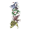
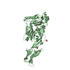
- PDB-4cyl: Tomographic subvolume average of EFF-1 fusogen on extracellular v... -
+ Open data

- Basic information
| Entry | Database: PDB / ID: 4cyl | ||||||
|---|---|---|---|---|---|---|---|
| Title | Tomographic subvolume average of EFF-1 fusogen on extracellular vesicles | ||||||
 Components Components | EFF-1A | ||||||
 Keywords Keywords | CELL ADHESION / CELL-CELL FUSION / EXTRACELLULAR FUSION / MEMBRANE FUSION / PRE-FUSION STATE | ||||||
| Function / homology |  Function and homology information Function and homology informationnematode male tail mating organ morphogenesis / fusogenic activity / nematode pharyngeal muscle development / EFF-1 complex / post-embryonic body morphogenesis / nematode male tail tip morphogenesis / cell-cell fusion / vulval development / syncytium formation by plasma membrane fusion / embryonic body morphogenesis ...nematode male tail mating organ morphogenesis / fusogenic activity / nematode pharyngeal muscle development / EFF-1 complex / post-embryonic body morphogenesis / nematode male tail tip morphogenesis / cell-cell fusion / vulval development / syncytium formation by plasma membrane fusion / embryonic body morphogenesis / egg-laying behavior / cell-cell contact zone / locomotion / morphogenesis of an epithelium / kinase activity / identical protein binding / plasma membrane / cytoplasm Similarity search - Function | ||||||
| Biological species |  | ||||||
| Method | ELECTRON MICROSCOPY / electron tomography / cryo EM / Resolution: 22.2 Å | ||||||
 Authors Authors | Zeev-Ben-Mordehai, T. / Vasishtan, D. / Siebert, C.A. / Grunewald, K. | ||||||
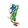
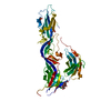
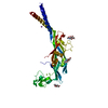
 Citation Citation | Journal: Nat Commun / Year: 2014 Title: The full-length cell-cell fusogen EFF-1 is monomeric and upright on the membrane. Authors: Tzviya Zeev-Ben-Mordehai / Daven Vasishtan / C Alistair Siebert / Kay Grünewald /  Abstract: Fusogens are membrane proteins that remodel lipid bilayers to facilitate membrane merging. Although several fusogen ectodomain structures have been solved, structural information on full-length, ...Fusogens are membrane proteins that remodel lipid bilayers to facilitate membrane merging. Although several fusogen ectodomain structures have been solved, structural information on full-length, natively membrane-anchored fusogens is scarce. Here we present the electron cryo microscopy three-dimensional reconstruction of the Caenorhabditis elegans epithelial fusion failure 1 (EFF-1) protein natively anchored in cell-derived membrane vesicles. This reveals a membrane protruding, asymmetric, elongated monomer. Flexible fitting of a protomer of the EFF-1 crystal structure, which is homologous to viral class-II fusion proteins, shows that EFF-1 has a hairpin monomeric conformation before fusion. These structural insights, when combined with our observations of membrane-merging intermediates between vesicles, enable us to propose a model for EFF-1 mediated fusion. This process, involving identical proteins on both membranes to be fused, follows a mechanism that shares features of SNARE-mediated fusion while using the structural building blocks of the unilaterally acting class-II viral fusion proteins. | ||||||
| History |
|
- Structure visualization
| Movie |
Movie viewer |
|---|---|
| Structure viewer | Molecule: MolmilJmol/JSmol |
- Downloads & links
-Download
| PDBx/mmCIF format | 4cyl.cif.gz | 100.4 KB | Display | PDBx/mmCIF format |
|---|---|---|---|---|
| PDB format | pdb4cyl.ent.gz | 78.2 KB | Display | PDB format |
| PDBx/mmJSON format | 4cyl.json.gz | Tree view | PDBx/mmJSON format | |
| Others |  Other downloads Other downloads |
-Validation report
| Summary document | 4cyl_validation.pdf.gz | 561.8 KB | Display | wwPDB validaton report |
|---|---|---|---|---|
| Full document | 4cyl_full_validation.pdf.gz | 579.5 KB | Display | |
| Data in XML | 4cyl_validation.xml.gz | 23.3 KB | Display | |
| Data in CIF | 4cyl_validation.cif.gz | 33.3 KB | Display | |
| Arichive directory | https://data.pdbj.org/pub/pdb/validation_reports/cy/4cylftp://data.pdbj.org/pub/pdb/validation_reports/cy/4cyl | HTTPS FTP |
-Related structure data
| Related structure data | 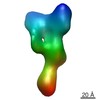 2530MC  2531C 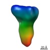 2532C M: map data used to model this data C: citing same article ( |
|---|---|
| Similar structure data |
-Links
 PDBj
PDBj- Assembly
| Deposited unit | 
|
|---|---|
| 1 |
|
-Components
| #1: Protein | Mass: 74497.141 Da / Num. of mol.: 1 Source method: isolated from a genetically manipulated source Details: PROTEINS ON EXTRACELLULAR VESICLES / Source: (gene. exp.)  MESOCRICETUS AURATUS (golden hamster) / References: UniProt: G5ECA1 MESOCRICETUS AURATUS (golden hamster) / References: UniProt: G5ECA1 |
|---|
-Experimental details
-Experiment
| Experiment | Method: ELECTRON MICROSCOPY |
|---|---|
| EM experiment | Aggregation state: PARTICLE / 3D reconstruction method: electron tomography |
- Sample preparation
| Component | Name: EPITHELIAL FUSION FAILURE 1 (EFF-1) ISOFORM A ON EXTRACELLULAR VESICLES Type: COMPLEX |
|---|---|
| Buffer solution | Name: 25MM HEPES, 130MM NACL / pH: 7.4 / Details: 25MM HEPES, 130MM NACL |
| Specimen | Embedding applied: NO / Shadowing applied: NO / Staining applied: NO / Vitrification applied: YES |
| Specimen support | Details: HOLEY CARBON |
| Vitrification | Instrument: HOMEMADE PLUNGER / Cryogen name: ETHANE-PROPANE Details: VITRIFICATION 1 -- CRYOGEN- ETHANE-PROPANE MIXTURE, TEMPERATURE- 77, INSTRUMENT- HOMEMADE PLUNGER, METHOD- BLOT FOR 3 SECONDS BEFORE PLUNGING |
- Electron microscopy imaging
| Experimental equipment |  Model: Tecnai F30 / Image courtesy: FEI Company |
|---|---|
| Microscopy | Model: FEI TECNAI F30 / Date: Mar 3, 2013 |
| Electron gun | Electron source:  FIELD EMISSION GUN / Accelerating voltage: 200 kV / Illumination mode: FLOOD BEAM FIELD EMISSION GUN / Accelerating voltage: 200 kV / Illumination mode: FLOOD BEAM |
| Electron lens | Mode: BRIGHT FIELD / Nominal magnification: 95000 X / Calibrated magnification: 78950 X / Nominal defocus max: 2500 nm / Nominal defocus min: 2000 nm / Cs: 2 mm |
| Specimen holder | Temperature: 85 K / Tilt angle max: 60 ° / Tilt angle min: -60 ° |
| Image recording | Electron dose: 80 e/Å2 / Film or detector model: GATAN ULTRASCAN 4000 (4k x 4k) |
| Image scans | Num. digital images: 78 |
| Radiation wavelength | Relative weight: 1 |
- Processing
| EM software |
| ||||||||||||
|---|---|---|---|---|---|---|---|---|---|---|---|---|---|
| Symmetry | Point symmetry: C1 (asymmetric) | ||||||||||||
| 3D reconstruction | Method: SUBTOMOGRAM AVERAGING / Resolution: 22.2 Å / Num. of particles: 801 / Nominal pixel size: 3.8 Å / Actual pixel size: 3.8 Å Details: SUBMISSION BASED ON EXPERIMENTAL DATA FROM EMDB EMD-2530. (DEPOSITION ID: 12175). Symmetry type: POINT | ||||||||||||
| Atomic model building | Protocol: FLEXIBLE FIT / Space: REAL Details: METHOD--FLEXIBLE FITTING REFINEMENT PROTOCOL--FLEXIBLE FIT OF X-RAY STRUCTURE | ||||||||||||
| Refinement | Highest resolution: 22.2 Å | ||||||||||||
| Refinement step | Cycle: LAST / Highest resolution: 22.2 Å
|
