 Movie
Movie Controller

[English] 日本語
 Yorodumi
- PDB-4ojc: Crystal structure of the wild-type full-length trimeric ectodomai... -
+ Open data

- Basic information
| Entry | Database: PDB / ID: 4ojc | |||||||||
|---|---|---|---|---|---|---|---|---|---|---|
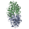
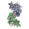
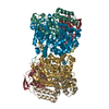
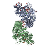
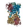
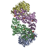
| Title | Crystal structure of the wild-type full-length trimeric ectodomain of the C. elegans fusion protein EFF-1 | |||||||||
 Components Components | EFF-1A | |||||||||
 Keywords Keywords |  MEMBRANE PROTEIN / class II fusion protein / membrane fusion protein / cell surface MEMBRANE PROTEIN / class II fusion protein / membrane fusion protein / cell surface | |||||||||
| Function / homology |  Function and homology information Function and homology informationnematode male tail mating organ morphogenesis / fusogenic activity / EFF-1 complex / chordate pharyngeal muscle development / post-embryonic body morphogenesis / nematode male tail tip morphogenesis / vulval development / cell-cell fusion / syncytium formation by plasma membrane fusion / embryonic body morphogenesis ...nematode male tail mating organ morphogenesis / fusogenic activity / EFF-1 complex / chordate pharyngeal muscle development / post-embryonic body morphogenesis / nematode male tail tip morphogenesis / vulval development / cell-cell fusion / syncytium formation by plasma membrane fusion / embryonic body morphogenesis / egg-laying behavior / cell-cell contact zone / locomotion / morphogenesis of an epithelium / kinase activity / phosphorylation / identical protein binding / plasma membrane / cytoplasmSimilarity search - Function | |||||||||
| Biological species |  Caenorhabditis elegans (invertebrata) Caenorhabditis elegans (invertebrata) | |||||||||
| Method | X-RAY DIFFRACTION / SYNCHROTRON / SAD / Resolution: 2.93 Å | |||||||||
 Authors Authors | Krey, T. / Rey, F.A. | |||||||||
 Citation Citation | Journal: Cell(Cambridge,Mass.) / Year: 2014 Title: Structural basis of eukaryotic cell-cell fusion Authors: Perez-Vargas, J. / Krey, T. / Valansi, C. / Avinoam, O. / Haouz, A. / Jamin, M. / Raveh-Barak, H. / Podbilewicz, B. / Rey, F.A. | |||||||||
| History |
|
- Structure visualization
| Structure viewer | Molecule: MolmilJmol/JSmol |
|---|
- Downloads & links
-Download
| PDBx/mmCIF format | 4ojc.cif.gz | 107.1 KB | Display | PDBx/mmCIF format |
|---|---|---|---|---|
| PDB format | pdb4ojc.ent.gz | 83.8 KB | Display | PDB format |
| PDBx/mmJSON format | 4ojc.json.gz | Tree view | PDBx/mmJSON format | |
| Others |  Other downloads Other downloads |
-Validation report
| Arichive directory | https://data.pdbj.org/pub/pdb/validation_reports/oj/4ojcftp://data.pdbj.org/pub/pdb/validation_reports/oj/4ojc | HTTPS FTP |
|---|
-Related structure data


-Links
 PDBj
PDBj- Assembly
| Deposited unit | 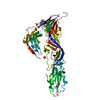
| ||||||||
|---|---|---|---|---|---|---|---|---|---|
| 1 | 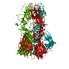
| ||||||||
| Unit cell |
| ||||||||
| Components on special symmetry positions |
|
-Components
| #1: Protein | Mass: 64947.637 Da / Num. of mol.: 1 / Fragment: UNP residues 23-561 Source method: isolated from a genetically manipulated source Source: (gene. exp.) Caenorhabditis elegans (invertebrata) / Gene: C26D10.5, CELE_C26D10.5, eff-1 / Plasmid: pMT-based / Cell (production host): S2 cells / Production host:  Drosophila melanogaster (fruit fly) / References: UniProt: G5ECA1 Drosophila melanogaster (fruit fly) / References: UniProt: G5ECA1 | ||||
|---|---|---|---|---|---|
| #2: Polysaccharide | 2-acetamido-2-deoxy-beta-D-glucopyranose-(1-4)-2-acetamido-2-deoxy-beta-D-glucopyranose / Mass: 424.401 Da / Num. of mol.: 1 Source method: isolated from a genetically manipulated source | ||||
| #3: Sugar | N-Acetylglucosamine  Type: D-saccharide, beta linking / Mass: 221.208 Da / Num. of mol.: 3 Type: D-saccharide, beta linking / Mass: 221.208 Da / Num. of mol.: 3Source method: isolated from a genetically manipulated source Formula: C8H15NO6 #4: Chemical | ChemComp-SO4 / Sulfate  Mass: 96.063 Da / Num. of mol.: 9 / Source method: obtained synthetically / Formula: SO4 Mass: 96.063 Da / Num. of mol.: 9 / Source method: obtained synthetically / Formula: SO4#5: Water | ChemComp-HOH / | Water Mass: 18.015 Da / Num. of mol.: 19 / Source method: isolated from a natural source / Formula: H2O Mass: 18.015 Da / Num. of mol.: 19 / Source method: isolated from a natural source / Formula: H2O |
-Experimental details
-Experiment
| Experiment | Method: X-RAY DIFFRACTION / Number of used crystals: 1 |
|---|
- Sample preparation
| Crystal | Density Matthews: 3.36 Å3/Da / Density % sol: 63.42 % |
|---|---|
| Crystal grow | Temperature: 293 K / Method: vapor diffusion, hanging drop / pH: 7 Details: 2% 2-Propanol, 50mM sodium citrate, 0.8-1.2M ammonium sulfate, pH 7.0, VAPOR DIFFUSION, HANGING DROP, temperature 293K |
-Data collection
| Diffraction | Mean temperature: 110 K |
|---|---|
| Diffraction source | Source: SYNCHROTRON / Site: SLS  / Beamline: X06SA / Wavelength: 1.0396 Å / Beamline: X06SA / Wavelength: 1.0396 Å |
| Detector | Type: PSI PILATUS 6M / Detector: PIXEL / Date: Mar 28, 2010 |
| Radiation | Monochromator: LN2 cooled Fixed-exit Si(111) / Protocol: SINGLE WAVELENGTH / Monochromatic (M) / Laue (L): M / Scattering type: x-ray |
| Radiation wavelength | Wavelength: 1.0396 Å / Relative weight: 1 |
| Reflection | Resolution: 2.93→50 Å / Num. all: 18909 / Num. obs: 18417 / % possible obs: 97.4 % / Observed criterion σ(F): 0 / Observed criterion σ(I): -3 / Redundancy: 7.2 % / Biso Wilson estimate: 113.4 Å2 |
| Reflection shell | Resolution: 2.93→3.09 Å / Redundancy: 4.2 % / Mean I/σ(I) obs: 3.1 / Num. unique all: 2735 |
- Processing
| Software |
| ||||||||||||||||||||||||||||||||||||||||||||||||||||||||||||||||||||||||||||||||||||||||||||||||||||||||||||||||||
|---|---|---|---|---|---|---|---|---|---|---|---|---|---|---|---|---|---|---|---|---|---|---|---|---|---|---|---|---|---|---|---|---|---|---|---|---|---|---|---|---|---|---|---|---|---|---|---|---|---|---|---|---|---|---|---|---|---|---|---|---|---|---|---|---|---|---|---|---|---|---|---|---|---|---|---|---|---|---|---|---|---|---|---|---|---|---|---|---|---|---|---|---|---|---|---|---|---|---|---|---|---|---|---|---|---|---|---|---|---|---|---|---|---|---|---|
| Refinement | Method to determine structure: SAD / Resolution: 2.93→46.42 Å / Cor.coef. Fo:Fc: 0.9231 / Cor.coef. Fo:Fc free: 0.8869 / SU R Cruickshank DPI: 0.814 / Cross valid method: THROUGHOUT / σ(F): 0 / Stereochemistry target values: Engh & Huber
| ||||||||||||||||||||||||||||||||||||||||||||||||||||||||||||||||||||||||||||||||||||||||||||||||||||||||||||||||||
| Displacement parameters | Biso mean: 92.38 Å2
| ||||||||||||||||||||||||||||||||||||||||||||||||||||||||||||||||||||||||||||||||||||||||||||||||||||||||||||||||||
| Refine analyze | Luzzati coordinate error obs: 0.549 Å | ||||||||||||||||||||||||||||||||||||||||||||||||||||||||||||||||||||||||||||||||||||||||||||||||||||||||||||||||||
| Refinement step | Cycle: LAST / Resolution: 2.93→46.42 Å
| ||||||||||||||||||||||||||||||||||||||||||||||||||||||||||||||||||||||||||||||||||||||||||||||||||||||||||||||||||
| Refine LS restraints |
| ||||||||||||||||||||||||||||||||||||||||||||||||||||||||||||||||||||||||||||||||||||||||||||||||||||||||||||||||||
| LS refinement shell | Resolution: 2.93→3.11 Å / Total num. of bins used: 9
|
