 Movie
Movie Controller


+ Open data

- Basic information
| Entry | Database: EMDB / ID: EMD-5124 | |||||||||
|---|---|---|---|---|---|---|---|---|---|---|
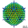
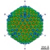
| Title | Cryo-EM of Rift Valley fever virus | |||||||||
 Map data Map data | this is RVFV map in 2-fold orientation | |||||||||
 Sample Sample |
| |||||||||
 Keywords Keywords | cryo-electron microscopy / single particle averaging / Rift valley fever / bunyaviridae / inter-capsomer contacts | |||||||||
| Biological species |  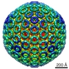 Rift Valley fever virus Rift Valley fever virus | |||||||||
| Method | single particle reconstruction / cryo EM / Resolution: 27.0 Å | |||||||||
 Authors Authors | Sherman MB / Freiberg AN / Holbrook AM / Watowich SJ | |||||||||
 Citation Citation | Journal: Virology / Year: 2009 Title: Single-particle cryo-electron microscopy of Rift Valley fever virus. Authors: Michael B Sherman / Alexander N Freiberg / Michael R Holbrook / Stanley J Watowich /  Abstract: Rift Valley fever virus (RVFV; Bunyaviridae; Phlebovirus) is an emerging human and veterinary pathogen causing acute hepatitis in ruminants and has the potential to cause hemorrhagic fever in humans. ...Rift Valley fever virus (RVFV; Bunyaviridae; Phlebovirus) is an emerging human and veterinary pathogen causing acute hepatitis in ruminants and has the potential to cause hemorrhagic fever in humans. We report a three-dimensional reconstruction of RVFV vaccine strain MP-12 (RVFV MP-12) by cryo-electron microcopy using icosahedral symmetry of individual virions. Although the genomic core of RVFV MP-12 is apparently poorly ordered, the glycoproteins on the virus surface are highly symmetric and arranged on a T=12 icosahedral lattice. Our RVFV MP-12 structure allowed clear identification of inter-capsomer contacts and definition of possible glycoprotein arrangements within capsomers. This structure provides a detailed model for phleboviruses, opens new avenues for high-resolution structural studies of the bunyavirus family, and aids the design of antiviral diagnostics and effective subunit vaccines. #1: Journal: J.VIROL. / Year: 2008Title: Three-dimensional organization of Rift Valley fever virus revealed by cryoelectron tomography Authors: Freiberg AN / Sherman MB / Morais MC / Holbrook MR / Watowich SJ | |||||||||
| History |
|
- Structure visualization
| Movie |
 Movie viewer Movie viewer |
|---|---|
| Structure viewer | EM map: SurfViewMolmilJmol/JSmol |
| Supplemental images |

- Downloads & links
-EMDB archive
| Map data | emd_5124.map.gz | 144.3 MB | EMDB map data format | |
|---|---|---|---|---|
| Header (meta data) | emd-5124-v30.xmlemd-5124.xml | 11.3 KB 11.3 KB | Display Display | EMDB header |
| Images | 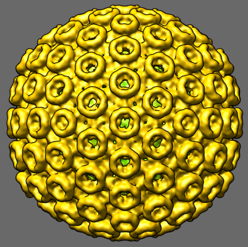 emd_5124_1.png emd_5124_1.png | 347.3 KB | ||
| Archive directory |  http://ftp.pdbj.org/pub/emdb/structures/EMD-5124ftp://ftp.pdbj.org/pub/emdb/structures/EMD-5124 http://ftp.pdbj.org/pub/emdb/structures/EMD-5124ftp://ftp.pdbj.org/pub/emdb/structures/EMD-5124 | HTTPS FTP |
-Validation report
| Summary document | emd_5124_validation.pdf.gz | 77.8 KB | Display | EMDB validaton report |
|---|---|---|---|---|
| Full document | emd_5124_full_validation.pdf.gz | 76.9 KB | Display | |
| Data in XML | emd_5124_validation.xml.gz | 493 B | Display | |
| Arichive directory | https://ftp.pdbj.org/pub/emdb/validation_reports/EMD-5124ftp://ftp.pdbj.org/pub/emdb/validation_reports/EMD-5124 | HTTPS FTP |
-Related structure data
| Similar structure data |
|---|
-Links
| EMDB pages | EMDB (EBI/PDBe) / EMDataResource |
|---|
-Map
| File | Download / File: emd_5124.map.gz / Format: CCP4 / Size: 341.7 MB / Type: IMAGE STORED AS FLOATING POINT NUMBER (4 BYTES) | ||||||||||||||||||||||||||||||||||||||||||||||||||||||||||||||||||||
|---|---|---|---|---|---|---|---|---|---|---|---|---|---|---|---|---|---|---|---|---|---|---|---|---|---|---|---|---|---|---|---|---|---|---|---|---|---|---|---|---|---|---|---|---|---|---|---|---|---|---|---|---|---|---|---|---|---|---|---|---|---|---|---|---|---|---|---|---|---|
| Annotation | this is RVFV map in 2-fold orientation | ||||||||||||||||||||||||||||||||||||||||||||||||||||||||||||||||||||
| Voxel size | X=Y=Z: 3 Å | ||||||||||||||||||||||||||||||||||||||||||||||||||||||||||||||||||||
| Density |
| ||||||||||||||||||||||||||||||||||||||||||||||||||||||||||||||||||||
| Symmetry | Space group: 1 | ||||||||||||||||||||||||||||||||||||||||||||||||||||||||||||||||||||
| Details | EMDB XML:
CCP4 map header:
| ||||||||||||||||||||||||||||||||||||||||||||||||||||||||||||||||||||
-Supplemental data
- Sample components
-Entire : Rift Valley fever virus, RVFV vaccine strain MP-12
| Entire | Name: Rift Valley fever virus, RVFV vaccine strain MP-12 |
|---|---|
| Components |
|
-Supramolecule #1000: Rift Valley fever virus, RVFV vaccine strain MP-12
| Supramolecule | Name: Rift Valley fever virus, RVFV vaccine strain MP-12 / type: sample / ID: 1000 / Details: monodisperse sample / Oligomeric state: single virions / Number unique components: 8 |
|---|
-Supramolecule #1: Rift Valley fever virus
| Supramolecule | Name: Rift Valley fever virus / type: virus / ID: 1 / Name.synonym: RVFV / Details: 100 nm particles / NCBI-ID: 11588 / Sci species name: Rift Valley fever virus / Database: NCBI / Virus type: VIRION / Virus isolate: STRAIN / Virus enveloped: Yes / Virus empty: No / Syn species name: RVFV |
|---|---|
| Host (natural) | Organism:  Homo sapiens (human) / synonym: VERTEBRATES Homo sapiens (human) / synonym: VERTEBRATES |
| Virus shell | Shell ID: 1 / Diameter: 1000 Å / T number (triangulation number): 12 |
-Experimental details
-Structure determination
| Method | cryo EM |
|---|---|
 Processing Processing | single particle reconstruction |
| Aggregation state | particle |
-Sample preparation
| Concentration | 0.1 mg/mL |
|---|---|
| Buffer | pH: 7.2 / Details: 10 mM Tris-HCl, 100 mM NaCl, 1 mM EDTA |
| Grid | Details: 200 mesh copper Qauntifoil grids |
| Vitrification | Cryogen name: ETHANE / Chamber humidity: 80 % / Chamber temperature: 95 K / Instrument: HOMEMADE PLUNGER / Details: Vitrification instrument: pneumatic plunger / Method: blot 2 sec before plunging |
- Electron microscopy
| Microscope | JEOL 2200FS |
|---|---|
| Temperature | Min: 99 K / Max: 99 K / Average: 99 K |
| Alignment procedure | Legacy - Astigmatism: objective lens astigmatism was corrected at 100,000 times magnification Legacy - Electron beam tilt params: 0 |
| Specialist optics | Energy filter - Name: omega / Energy filter - Lower energy threshold: 0.0 eV / Energy filter - Upper energy threshold: 20.0 eV |
| Date | Nov 1, 2007 |
| Image recording | Category: CCD / Film or detector model: GATAN ULTRASCAN 4000 (4k x 4k) / Digitization - Sampling interval: 14 µm / Number real images: 93 / Average electron dose: 20 e/Å2 / Bits/pixel: 16 |
| Tilt angle min | 0 |
| Tilt angle max | 0 |
| Electron beam | Acceleration voltage: 200 kV / Electron source:  FIELD EMISSION GUN FIELD EMISSION GUN |
| Electron optics | Calibrated magnification: 40200 / Illumination mode: FLOOD BEAM / Imaging mode: BRIGHT FIELD / Cs: 2.0 mm / Nominal defocus max: 3.7 µm / Nominal defocus min: 0.7 µm / Nominal magnification: 40000 |
| Sample stage | Specimen holder: Eucentric / Specimen holder model: GATAN LIQUID NITROGEN |
-Image processing
| CTF correction | Details: Each particle |
|---|---|
| Final reconstruction | Algorithm: OTHER / Resolution.type: BY AUTHOR / Resolution: 27.0 Å / Resolution method: OTHER / Software - Name: PFT / Details: Final map calculated from 108 particles / Number images used: 108 |
| Final two d classification | Number classes: 1 |
