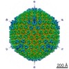
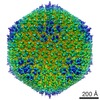
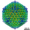
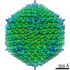
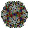
Journal: Structure / Year: 2019 Title: Capsid Structure of a Freshwater Cyanophage Siphoviridae Mic1. Authors: Hua Jin / Yong-Liang Jiang / Feng Yang / Jun-Tao Zhang / Wei-Fang Li / Ke Zhou / Jue Ju / Yuxing Chen / Cong-Zhao Zhou / Abstract: Cyanobacteria are the most abundant photosynthetic microorganisms, the global distribution of which is mainly regulated by the corresponding cyanophages. A systematic screening of water samples in ...Cyanobacteria are the most abundant photosynthetic microorganisms, the global distribution of which is mainly regulated by the corresponding cyanophages. A systematic screening of water samples in the Lake Chaohu enabled us to isolate a freshwater siphocyanophage that infects Microcystis wesenbergii, thus termed Mic1. Using cryoelectron microscopy, we solved the 3.5-Å structure of Mic1 capsid. The major capsid protein gp40 of an HK97-like fold forms two types of capsomers, hexons and pentons. The capsomers interact with each other via the interweaved N-terminal arms of gp40 in addition to a tail-in-mouth joint along the three-fold symmetric axis, resulting in the assembly of capsid in a mortise-and-tenon pattern. The novel-fold cement protein gp47 sticks at the two-fold symmetric axis and further fixes the capsid. These findings provide structural insights into the assembly of cyanophages, and set up a platform to explore the mechanism of specific interactions and co-evolution with cyanobacteria.
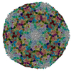
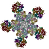
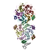
A: major capsid protein B: major capsid protein C: major capsid protein D: major capsid protein E: major capsid protein F: major capsid protein U: major capsid protein V: major capsid protein W: major capsid protein X: major capsid protein Y: major capsid protein Z: major capsid protein a: major capsid protein 0: cement protein 3: cement protein 1: cement protein 4: cement protein 2: cement protein 6: cement protein 5: cement protein 7: cement protein b: cement protein 8: cement protein c: cement protein 9: cement protein d: cement protein
A: major capsid protein B: major capsid protein C: major capsid protein D: major capsid protein E: major capsid protein F: major capsid protein U: major capsid protein V: major capsid protein W: major capsid protein X: major capsid protein Y: major capsid protein Z: major capsid protein a: major capsid protein 0: cement protein 3: cement protein 1: cement protein 4: cement protein 2: cement protein 6: cement protein 5: cement protein 7: cement protein b: cement protein 8: cement protein c: cement protein 9: cement protein d: cement protein
A: major capsid protein B: major capsid protein C: major capsid protein D: major capsid protein E: major capsid protein F: major capsid protein U: major capsid protein V: major capsid protein W: major capsid protein X: major capsid protein Y: major capsid protein Z: major capsid protein a: major capsid protein 0: cement protein 3: cement protein 1: cement protein 4: cement protein 2: cement protein 6: cement protein 5: cement protein 7: cement protein b: cement protein 8: cement protein c: cement protein 9: cement protein d: cement protein
x 5
icosahedral pentamer
3.13 MDa, 130 polymers
Theoretical mass
Number of molelcules
Total (without water)
3,125,102
130
Polymers
3,125,102
130
Non-polymers
0
0
Water
0
Type
Name
Symmetry operation
Number
identity operation
1_555
x,y,z
1
point symmetry operation
4
4
A: major capsid protein B: major capsid protein C: major capsid protein D: major capsid protein E: major capsid protein F: major capsid protein U: major capsid protein V: major capsid protein W: major capsid protein X: major capsid protein Y: major capsid protein Z: major capsid protein a: major capsid protein 0: cement protein 3: cement protein 1: cement protein 4: cement protein 2: cement protein 6: cement protein 5: cement protein 7: cement protein b: cement protein 8: cement protein c: cement protein 9: cement protein d: cement protein
x 6
icosahedral 23 hexamer
3.75 MDa, 156 polymers
Theoretical mass
Number of molelcules
Total (without water)
3,750,122
156
Polymers
3,750,122
156
Non-polymers
0
0
Water
0
Type
Name
Symmetry operation
Number
identity operation
1_555
x,y,z
1
point symmetry operation
5
5
Idetical with deposited unit in distinct coordinate
icosahedral asymmetric unit, std point frame
Type
Name
Symmetry operation
Number
transform to point frame
1
Symmetry
Point symmetry: (Schoenflies symbol: I (icosahedral))
-
Components
#1: Protein
majorcapsidprotein
Mass: 37879.035 Da / Num. of mol.: 13 / Source method: isolated from a natural source / Source: (natural) Microcystis phage Mic1 (virus) / References: UniProt: A0A4Y5TR23*PLUS
#2: Protein
cementprotein
Mass: 10199.458 Da / Num. of mol.: 13 / Source method: isolated from a natural source / Source: (natural) Microcystis phage Mic1 (virus) / References: UniProt: A0A4Y5TPY8*PLUS
-
Experimental details
-
Experiment
Experiment
Method: ELECTRON MICROSCOPY
EM experiment
Aggregation state: PARTICLE / 3D reconstruction method: single particle reconstruction
Average exposure time: 5.76 sec. / Electron dose: 25 e/Å2 / Detector mode: SUPER-RESOLUTION / Film or detector model: GATAN K2 SUMMIT (4k x 4k) / Num. of grids imaged: 1 / Num. of real images: 1935
Image scans
Movie frames/image: 32
-
Processing
EM software
ID
Name
Version
Category
2
SerialEM
imageacquisition
4
CTFFIND
CTFcorrection
7
UCSF Chimera
modelfitting
9
PHENIX
modelrefinement
10
RELION
2
initialEulerassignment
11
RELION
2
finalEulerassignment
12
RELION
2
classification
13
RELION
2
3Dreconstruction
CTF correction
Details: CTFFIND4 / Type: NONE
Particle selection
Num. of particles selected: 18800
Symmetry
Point symmetry: I (icosahedral)
3D reconstruction
Resolution: 3.53 Å / Resolution method: FSC 0.143 CUT-OFF / Num. of particles: 9702 / Num. of class averages: 1 / Symmetry type: POINT
Atomic model building
Protocol: AB INITIO MODEL
+
About Yorodumi
-
News
-
Feb 9, 2022. New format data for meta-information of EMDB entries
New format data for meta-information of EMDB entries
Version 3 of the EMDB header file is now the official format.
The previous official version 1.9 will be removed from the archive.
In the structure databanks used in Yorodumi, some data are registered as the other names, "COVID-19 virus" and "2019-nCoV". Here are the details of the virus and the list of structure data.
Jan 31, 2019. EMDB accession codes are about to change! (news from PDBe EMDB page)
EMDB accession codes are about to change! (news from PDBe EMDB page)
The allocation of 4 digits for EMDB accession codes will soon come to an end. Whilst these codes will remain in use, new EMDB accession codes will include an additional digit and will expand incrementally as the available range of codes is exhausted. The current 4-digit format prefixed with “EMD-” (i.e. EMD-XXXX) will advance to a 5-digit format (i.e. EMD-XXXXX), and so on. It is currently estimated that the 4-digit codes will be depleted around Spring 2019, at which point the 5-digit format will come into force.
The EM Navigator/Yorodumi systems omit the EMD- prefix.
Related info.:Q: What is EMD? / ID/Accession-code notation in Yorodumi/EM Navigator
Yorodumi is a browser for structure data from EMDB, PDB, SASBDB, etc.
This page is also the successor to EM Navigator detail page, and also detail information page/front-end page for Omokage search.
The word "yorodu" (or yorozu) is an old Japanese word meaning "ten thousand". "mi" (miru) is to see.
Related info.:EMDB / PDB / SASBDB / Comparison of 3 databanks / Yorodumi Search / Aug 31, 2016. New EM Navigator & Yorodumi / Yorodumi Papers / Jmol/JSmol / Function and homology information / Changes in new EM Navigator and Yorodumi
 Movie
Movie Controller


 Open data
 Basic information
 Components
 Keywords
 VIRUS /
  Function and homology information
 Microcystis phage Mic1 (virus)
 Authors
 China, 4items
  Citation
 Structure visualization
 Downloads & links
 Other downloads
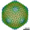


 PDBj
PDBj Assembly

 Sample preparation
 Electron microscopy imaging
 Processing