 Movie
Movie Controller

[English] 日本語
 Yorodumi
- EMDB-2978: Time-resolved Cryo Electron Microscopy of ribosome subunit association -
+ Open data

- Basic information
| Entry | Database: EMDB / ID: EMD-2978 | |||||||||
|---|---|---|---|---|---|---|---|---|---|---|
| Title | Time-resolved Cryo Electron Microscopy of ribosome subunit association | |||||||||
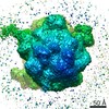
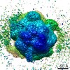
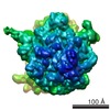
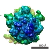
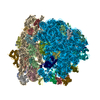
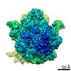
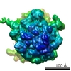
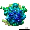
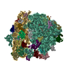
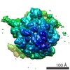
 Map data Map data | Reconstruction of E. Coli naked 70S ribosome in rotated (RT) conformation | |||||||||
 Sample Sample |
| |||||||||
 Keywords Keywords | time-resolved / cryo-EM / mixing-spraying / ribosome subunit association / structural dynamics | |||||||||
| Function / homology |  Function and homology information Function and homology informationCajal-Retzius cell differentiation / positive regulation of L-glutamate import across plasma membrane / amyloid precursor protein biosynthetic process / positive regulation of coagulation / negative regulation of core promoter binding / gamma-secretase complex / aspartic endopeptidase activity, intramembrane cleaving / short-term synaptic potentiation / protein catabolic process at postsynapse / positive regulation of amyloid precursor protein biosynthetic process ...Cajal-Retzius cell differentiation / positive regulation of L-glutamate import across plasma membrane / amyloid precursor protein biosynthetic process / positive regulation of coagulation / negative regulation of core promoter binding / gamma-secretase complex / aspartic endopeptidase activity, intramembrane cleaving / short-term synaptic potentiation / protein catabolic process at postsynapse / positive regulation of amyloid precursor protein biosynthetic process / positive regulation of endopeptidase activity / Noncanonical activation of NOTCH3 / sequestering of calcium ion / Notch receptor processing / choline transport / central nervous system myelination / synaptic vesicle targeting / membrane protein intracellular domain proteolysis / negative regulation of axonogenesis / regulation of resting membrane potential / T cell activation involved in immune response / NOTCH4 Activation and Transmission of Signal to the Nucleus / skin morphogenesis / growth factor receptor binding / regulation of synaptic vesicle cycle / dorsal/ventral neural tube patterning / neural retina development / L-glutamate import across plasma membrane / myeloid dendritic cell differentiation / Regulated proteolysis of p75NTR / cerebral cortex cell migration / regulation of phosphorylation / brain morphogenesis / endoplasmic reticulum calcium ion homeostasis / nuclear outer membrane / glutamate receptor signaling pathway / locomotion / amyloid precursor protein metabolic process / smooth endoplasmic reticulum calcium ion homeostasis / regulation of canonical Wnt signaling pathway / astrocyte activation involved in immune response / aggresome / regulation of long-term synaptic potentiation / embryonic limb morphogenesis / skeletal system morphogenesis / cell fate specification / ciliary rootlet / myeloid cell homeostasis / regulation of postsynapse organization / azurophil granule membrane / G protein-coupled dopamine receptor signaling pathway / Hydrolases; Acting on peptide bonds (peptidases); Aspartic endopeptidases / positive regulation of amyloid fibril formation / adult behavior / mitochondrial transport / positive regulation of dendritic spine development / positive regulation of receptor recycling / blood vessel development / regulation of neuron projection development / heart looping / protein glycosylation / amyloid precursor protein catabolic process / amyloid-beta formation / negative regulation of apoptotic signaling pathway / membrane protein ectodomain proteolysis / autophagosome assembly / EPH-ephrin mediated repulsion of cells / smooth endoplasmic reticulum / neuron development / hematopoietic progenitor cell differentiation / negative regulation of ubiquitin-dependent protein catabolic process / somitogenesis / calcium ion homeostasis / T cell proliferation / Nuclear signaling by ERBB4 / rough endoplasmic reticulum / viral release from host cell by cytolysis / epithelial cell proliferation / Notch signaling pathway / regulation of synaptic transmission, glutamatergic / neuron projection maintenance / NOTCH2 Activation and Transmission of Signal to the Nucleus / cellular response to calcium ion / astrocyte activation / NOTCH3 Activation and Transmission of Signal to the Nucleus / Degradation of the extracellular matrix / positive regulation of glycolytic process / NRIF signals cell death from the nucleus / peptidoglycan catabolic process / Activated NOTCH1 Transmits Signal to the Nucleus / cerebellum development / post-embryonic development / thymus development / negative regulation of protein phosphorylation / dendritic shaft / PDZ domain binding / apoptotic signaling pathway / cell-cell adhesion / synapse organization / neuron migration Similarity search - Function | |||||||||
| Biological species |  | |||||||||
| Method | single particle reconstruction / cryo EM / Resolution: 11.6 Å | |||||||||
 Authors Authors | Chen B / Kaledhonkar S / Sun M / Shen B / Lu Z / Barnard D / Lu T / Gonzalez Jr R / Frank J | |||||||||
 Citation Citation | Journal: Structure / Year: 2015 Title: Structural dynamics of ribosome subunit association studied by mixing-spraying time-resolved cryogenic electron microscopy. Authors: Bo Chen / Sandip Kaledhonkar / Ming Sun / Bingxin Shen / Zonghuan Lu / David Barnard / Toh-Ming Lu / Ruben L Gonzalez / Joachim Frank /  Abstract: Ribosomal subunit association is a key checkpoint in translation initiation but its structural dynamics are poorly understood. Here, we used a recently developed mixing-spraying, time-resolved, ...Ribosomal subunit association is a key checkpoint in translation initiation but its structural dynamics are poorly understood. Here, we used a recently developed mixing-spraying, time-resolved, cryogenic electron microscopy (cryo-EM) method to study ribosomal subunit association in the sub-second time range. We have improved this method and increased the cryo-EM data yield by tenfold. Pre-equilibrium states of the association reaction were captured by reacting the mixture of ribosomal subunits for 60 ms and 140 ms. We also identified three distinct ribosome conformations in the associated ribosomes. The observed proportions of these conformations are the same in these two time points, suggesting that ribosomes equilibrate among the three conformations within less than 60 ms upon formation. Our results demonstrate that the mixing-spraying method can capture multiple states of macromolecules during a sub-second reaction. Other fast processes, such as translation initiation, decoding, and ribosome recycling, are amenable to study with this method. | |||||||||
| History |
|
- Structure visualization
| Movie |
Movie viewer |
|---|---|
| Structure viewer | EM map: SurfViewMolmilJmol/JSmol |
| Supplemental images |

- Downloads & links
-EMDB archive
| Map data | emd_2978.map.gz | 14.3 MB | EMDB map data format | |
|---|---|---|---|---|
| Header (meta data) | emd-2978-v30.xmlemd-2978.xml | 9.2 KB 9.2 KB | Display Display | EMDB header |
| Images | 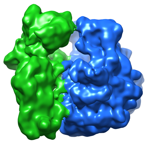 emd_2978.png emd_2978.png | 281.5 KB | ||
| Archive directory |  http://ftp.pdbj.org/pub/emdb/structures/EMD-2978ftp://ftp.pdbj.org/pub/emdb/structures/EMD-2978 http://ftp.pdbj.org/pub/emdb/structures/EMD-2978ftp://ftp.pdbj.org/pub/emdb/structures/EMD-2978 | HTTPS FTP |
-Validation report
| Summary document | emd_2978_validation.pdf.gz | 226 KB | Display | EMDB validaton report |
|---|---|---|---|---|
| Full document | emd_2978_full_validation.pdf.gz | 225.2 KB | Display | |
| Data in XML | emd_2978_validation.xml.gz | 5.4 KB | Display | |
| Arichive directory | https://ftp.pdbj.org/pub/emdb/validation_reports/EMD-2978ftp://ftp.pdbj.org/pub/emdb/validation_reports/EMD-2978 | HTTPS FTP |
-Related structure data
-Links
| EMDB pages | EMDB (EBI/PDBe) / EMDataResource |
|---|---|
| Related items in Molecule of the Month |


-Map
| File | Download / File: emd_2978.map.gz / Format: CCP4 / Size: 15.3 MB / Type: IMAGE STORED AS FLOATING POINT NUMBER (4 BYTES) | ||||||||||||||||||||||||||||||||||||||||||||||||||||||||||||
|---|---|---|---|---|---|---|---|---|---|---|---|---|---|---|---|---|---|---|---|---|---|---|---|---|---|---|---|---|---|---|---|---|---|---|---|---|---|---|---|---|---|---|---|---|---|---|---|---|---|---|---|---|---|---|---|---|---|---|---|---|---|
| Annotation | Reconstruction of E. Coli naked 70S ribosome in rotated (RT) conformation | ||||||||||||||||||||||||||||||||||||||||||||||||||||||||||||
| Voxel size | X=Y=Z: 2.2451 Å | ||||||||||||||||||||||||||||||||||||||||||||||||||||||||||||
| Density |
| ||||||||||||||||||||||||||||||||||||||||||||||||||||||||||||
| Symmetry | Space group: 1 | ||||||||||||||||||||||||||||||||||||||||||||||||||||||||||||
| Details | EMDB XML:
CCP4 map header:
| ||||||||||||||||||||||||||||||||||||||||||||||||||||||||||||
-Supplemental data
- Sample components
-Entire : E. Coli 70S Ribosome
| Entire | Name: E. Coli 70S Ribosome |
|---|---|
| Components |
|
-Supramolecule #1000: E. Coli 70S Ribosome
| Supramolecule | Name: E. Coli 70S Ribosome / type: sample / ID: 1000 / Number unique components: 1 |
|---|
-Supramolecule #1: 70S ribosome
| Supramolecule | Name: 70S ribosome / type: complex / ID: 1 / Recombinant expression: No / Ribosome-details: ribosome-prokaryote: LSU 50S, SSU 30S |
|---|---|
| Source (natural) | Organism: |
-Experimental details
-Structure determination
| Method | cryo EM |
|---|---|
 Processing Processing | single particle reconstruction |
| Aggregation state | particle |
-Sample preparation
| Buffer | pH: 7.6 Details: 25 mM Tris-HCl, 60 mM NH4Cl, 5 mM 2-mercaptoethanol, 3.5 mM MgCl2 |
|---|---|
| Grid | Details: Quantifoil R2/2 300 mesh copper grid with thin carbon sipport |
| Vitrification | Cryogen name: ETHANE / Chamber humidity: 80 % / Chamber temperature: 80 K / Instrument: OTHER Details: Equal volume of 1.2 microM 30S and 0.6 microM 50S (final concentration after mixing) were injected into the mixing-spraying device each at flow rate of 3 microL/s. The computer-controlled ...Details: Equal volume of 1.2 microM 30S and 0.6 microM 50S (final concentration after mixing) were injected into the mixing-spraying device each at flow rate of 3 microL/s. The computer-controlled plunging device was purchased from Dr. Howard White (Eastern Virginia Medical School, VA). Timed resolved state: Vitrified after spraying |
- Electron microscopy
| Microscope | FEI TECNAI F20 |
|---|---|
| Temperature | Average: 80 K |
| Details | Low dose, Data was collected over two years time |
| Date | Sep 13, 2013 |
| Image recording | Category: CCD / Film or detector model: GATAN ULTRASCAN 4000 (4k x 4k) / Number real images: 3402 / Average electron dose: 17 e/Å2 |
| Electron beam | Acceleration voltage: 200 kV / Electron source:  FIELD EMISSION GUN FIELD EMISSION GUN |
| Electron optics | Calibrated magnification: 66318 / Illumination mode: FLOOD BEAM / Imaging mode: BRIGHT FIELD / Cs: 2 mm / Nominal defocus max: 4.0 µm / Nominal defocus min: 2.0 µm / Nominal magnification: 50000 |
| Sample stage | Specimen holder: CT 3500 / Specimen holder model: GATAN LIQUID NITROGEN |
| Experimental equipment |  Model: Tecnai F20 / Image courtesy: FEI Company |
-Image processing
| Details | The partciles were selected with Autopicker (Langlois et al., 2014), and 3D classification and reconstruction with RELION |
|---|---|
| CTF correction | Details: each Micrograph |
| Final reconstruction | Applied symmetry - Point group: C1 (asymmetric) / Resolution.type: BY AUTHOR / Resolution: 11.6 Å / Resolution method: OTHER / Software - Name: Arachnid, RELION, SPIDER / Number images used: 11129 |
