Movie
Movie Controller

[English] 日本語
 Yorodumi
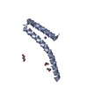
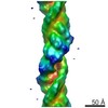
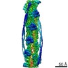
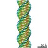
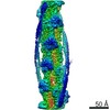
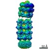
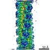
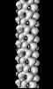
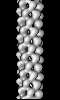
- EMDB-1416: Helical structure of the needle of the type III secretion system ... -
+ Open data

- Basic information
| Entry | Database: EMDB / ID: EMD-1416 | |||||||||
|---|---|---|---|---|---|---|---|---|---|---|
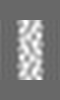
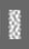
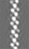
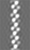
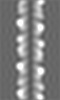
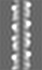
| Title | Helical structure of the needle of the type III secretion system of Shigella flexneri. | |||||||||
 Map data Map data | ccp4 map of reconstruction of Shigella flexneri T3SS needle | |||||||||
 Sample Sample |
| |||||||||
| Function / homology |  Function and homology information Function and homology informationtype III protein secretion system complex / protein secretion by the type III secretion system / cell surface / extracellular region / identical protein binding Similarity search - Function | |||||||||
| Biological species |  Shigella flexneri (bacteria) Shigella flexneri (bacteria) | |||||||||
| Method | helical reconstruction / cryo EM / negative staining / Resolution: 16.0 Å | |||||||||
 Authors Authors | Cordes FS / Komoriya K / Larquet E / Yang S / Egelman EH / Blocker A / Lea SM | |||||||||
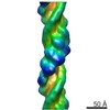
 Citation Citation | Journal: J Biol Chem / Year: 2003 Title: Helical structure of the needle of the type III secretion system of Shigella flexneri. Authors: Frank S Cordes / Kaoru Komoriya / Eric Larquet / Shixin Yang / Edward H Egelman / Ariel Blocker / Susan M Lea /  Abstract: Gram-negative bacteria commonly interact with animal and plant hosts using type III secretion systems (TTSSs) for translocation of proteins into eukaryotic cells during infection. 10 of the 25 TTSS- ...Gram-negative bacteria commonly interact with animal and plant hosts using type III secretion systems (TTSSs) for translocation of proteins into eukaryotic cells during infection. 10 of the 25 TTSS-encoding genes are homologous to components of the bacterial flagellar basal body, which the TTSS needle complex morphologically resembles. This indicates a common ancestry, although no TTSS sequence homologues for the genes encoding the flagellum are found. We here present an approximately 16-A structure of the central component, the needle, of the TTSS. Although the needle subunit is significantly smaller and shares no sequence homology with the flagellar hook and filament, it shares a common helical architecture ( approximately 5.6 subunits/turn, 24-A helical pitch). This common architecture implies that there will be further mechanistic analogies in the functioning of these two bacterial systems. | |||||||||
| History |
|
- Structure visualization
| Movie |
Movie viewer |
|---|---|
| Structure viewer | EM map: SurfViewMolmilJmol/JSmol |
| Supplemental images |
 UCSF Chimera
- Downloads & links
-EMDB archive
| Map data | emd_1416.map.gz | 281.1 KB | EMDB map data format | |
|---|---|---|---|---|
| Header (meta data) | emd-1416-v30.xmlemd-1416.xml | 10.3 KB 10.3 KB | Display Display | EMDB header |
| Images | 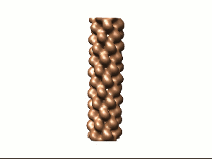 1416.gif 1416.gif | 10.9 KB | ||
| Archive directory |  http://ftp.pdbj.org/pub/emdb/structures/EMD-1416ftp://ftp.pdbj.org/pub/emdb/structures/EMD-1416 http://ftp.pdbj.org/pub/emdb/structures/EMD-1416ftp://ftp.pdbj.org/pub/emdb/structures/EMD-1416 | HTTPS FTP |
-Validation report
| Summary document | emd_1416_validation.pdf.gz | 197.2 KB | Display | EMDB validaton report |
|---|---|---|---|---|
| Full document | emd_1416_full_validation.pdf.gz | 196.3 KB | Display | |
| Data in XML | emd_1416_validation.xml.gz | 4.7 KB | Display | |
| Arichive directory | https://ftp.pdbj.org/pub/emdb/validation_reports/EMD-1416ftp://ftp.pdbj.org/pub/emdb/validation_reports/EMD-1416 | HTTPS FTP |
-Related structure data
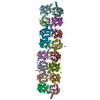
| Related structure data |  2v6lM M: atomic model generated by this map |
|---|---|
| Similar structure data |
-Links
| EMDB pages | EMDB (EBI/PDBe) / EMDataResource |
|---|
-Map
| File | Download / File: emd_1416.map.gz / Format: CCP4 / Size: 1.3 MB / Type: IMAGE STORED AS FLOATING POINT NUMBER (4 BYTES) | ||||||||||||||||||||||||||||||||||||||||||||||||||||||||||||||||||||
|---|---|---|---|---|---|---|---|---|---|---|---|---|---|---|---|---|---|---|---|---|---|---|---|---|---|---|---|---|---|---|---|---|---|---|---|---|---|---|---|---|---|---|---|---|---|---|---|---|---|---|---|---|---|---|---|---|---|---|---|---|---|---|---|---|---|---|---|---|---|
| Annotation | ccp4 map of reconstruction of Shigella flexneri T3SS needle | ||||||||||||||||||||||||||||||||||||||||||||||||||||||||||||||||||||
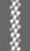
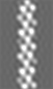
| Projections & slices | Image control
Images are generated by Spider. generated in cubic-lattice coordinate | ||||||||||||||||||||||||||||||||||||||||||||||||||||||||||||||||||||
| Voxel size | X=Y=Z: 2.65 Å | ||||||||||||||||||||||||||||||||||||||||||||||||||||||||||||||||||||
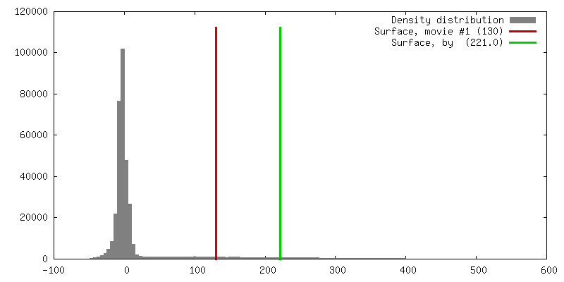
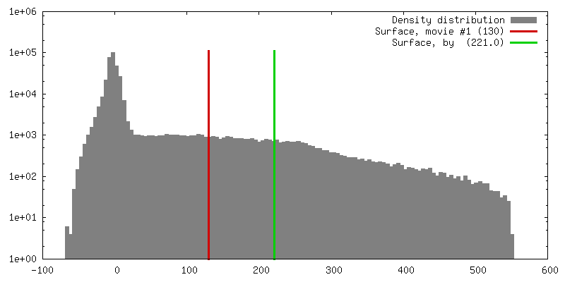
| Density |
| ||||||||||||||||||||||||||||||||||||||||||||||||||||||||||||||||||||
| Symmetry | Space group: 1 | ||||||||||||||||||||||||||||||||||||||||||||||||||||||||||||||||||||
| Details | EMDB XML:
CCP4 map header:
| ||||||||||||||||||||||||||||||||||||||||||||||||||||||||||||||||||||
 Z (Sec.)
 X (Row.)
 Y (Col.)


-Supplemental data
- Sample components
-Entire : Shigella flexneri T3SS needle - MxiH polymer
| Entire | Name: Shigella flexneri T3SS needle - MxiH polymer |
|---|---|
| Components |
|
-Supramolecule #1000: Shigella flexneri T3SS needle - MxiH polymer
| Supramolecule | Name: Shigella flexneri T3SS needle - MxiH polymer / type: sample / ID: 1000 / Oligomeric state: helical / Number unique components: 1 |
|---|
-Macromolecule #1: MxiH
| Macromolecule | Name: MxiH / type: protein_or_peptide / ID: 1 / Name.synonym: MxiH / Oligomeric state: helical polymer / Recombinant expression: Yes |
|---|---|
| Source (natural) | Organism: Shigella flexneri (bacteria) / Strain: M90T / Location in cell: extracellular |
| Molecular weight | Experimental: 9265 MDa |
| Recombinant expression | Organism: Shigella flexneri (bacteria) / Recombinant plasmid: pKT001 |
| Sequence | InterPro: Type III secretion system, needle protein |
-Experimental details
-Structure determination
| Method | negative staining, cryo EM |
|---|---|
 Processing Processing | helical reconstruction |
| Aggregation state | filament |
-Sample preparation
| Concentration | 0.1 mg/mL |
|---|---|
| Buffer | pH: 7.5 / Details: 10mM Tris pH 7.5 |
| Staining | Type: NEGATIVE / Details: 2% uranyl acetate, pH 7.5 |
| Grid | Details: 200 mesh carbon coated copper |
| Vitrification | Cryogen name: ETHANE |
- Electron microscopy
| Microscope | FEI/PHILIPS CM120T |
|---|---|
| Temperature | Average: 293 K |
| Details | low dose conditions |
| Image recording | Category: FILM / Film or detector model: KODAK SO-163 FILM / Digitization - Scanner: OTHER / Number real images: 18 / Average electron dose: 10 e/Å2 / Details: 1600dpi, 2.65 Angstrom per pixel |
| Tilt angle min | 0 |
| Tilt angle max | 0 |
| Electron beam | Acceleration voltage: 120 kV / Electron source: LAB6 |
| Electron optics | Illumination mode: SPOT SCAN / Imaging mode: BRIGHT FIELD / Cs: 2 mm / Nominal defocus max: 0.7 µm / Nominal defocus min: 0.7 µm / Nominal magnification: 60000 |
| Sample stage | Specimen holder: Eucentric / Specimen holder model: OTHER |
-Image processing
| Details | the 5-start helix is left handed |
|---|---|
| Final reconstruction | Algorithm: OTHER / Resolution.type: BY AUTHOR / Resolution: 16.0 Å / Resolution method: OTHER / Software - Name: SPIDER and MRC / Details: see paper |
-Atomic model buiding 1
| Initial model | PDB ID: |
|---|---|
| Software | Name: BUSTER-TnT |
| Details | Protocol: Rigid Body. Fitted by hand and optimised using URO. Helical parameters used. |
| Refinement | Space: RECIPROCAL / Protocol: RIGID BODY FIT / Overall B value: 50 / Target criteria: Correlation Coefficient and R_factor |
| Output model | PDB-2v6l: |