 Movie
Movie Controller


+ Open data

- Basic information
| Entry | Database: EMDB / ID: EMD-0786 | |||||||||
|---|---|---|---|---|---|---|---|---|---|---|
| Title | Cryo-EM Structure of YebT in conformation 1 | |||||||||
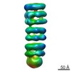
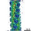
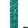
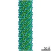
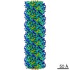
 Map data Map data | The whole map of YebT | |||||||||
 Sample Sample |
| |||||||||
| Biological species |   Escherichia coli K-12 (bacteria) Escherichia coli K-12 (bacteria) | |||||||||
| Method | single particle reconstruction / cryo EM / Resolution: 4.6 Å | |||||||||
 Authors Authors | Liu C / Zhang L / Wang HW / Wang J / Li XM | |||||||||
| Funding support |  China, 2 items China, 2 items
| |||||||||
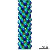
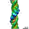
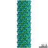
 Citation Citation | Journal: J Mol Biol / Year: 2020 Title: Cryo-EM Structure of a Bacterial Lipid Transporter YebT. Authors: Chuan Liu / Jinying Ma / Jia Wang / Hongwei Wang / Li Zhang / Abstract: The outer membrane (OM) of Gram-negative bacteria is asymmetric, with lipopolysaccharides (LPSs) on the outer surface and phospholipids (PLs) on the inner surface. This unique organization of OM ...The outer membrane (OM) of Gram-negative bacteria is asymmetric, with lipopolysaccharides (LPSs) on the outer surface and phospholipids (PLs) on the inner surface. This unique organization of OM makes Gram-negative bacteria resistant to many toxic chemicals. How this asymmetric distribution of lipids is maintained has been studied for decades with previous reports of an Mla (Maintenance of OM Lipid Asymmetry) system to be involved. Furthermore, the OM of Gram-negative bacteria is about 20 nm away from inner membrane (IM) where the lipids are synthesized. Therefore, how nascent lipids travel across the periplasmic space and arrive at the inner surface of OM is another interesting question. YebT is a homologue of MlaD in the Mla pathway, but its role in lipid distribution of the OM and IM is largely unknown. Here we report the first high-resolution (~3.0 Å) cryo-EM structure of full-length E. coli YebT in a substrate-bound state. Our structure with details of lipid interaction indicates that YebT is a lipid transporter spanning between IM and OM. We also demonstrate the symmetry mismatch in YebT and the existence of many other conformations of YebT revealing the intrinsic dynamics of this lipid channel. And a brief discussion on possible mechanisms of lipid transport is also included. | |||||||||
| History |
|
- Structure visualization
| Movie |
 Movie viewer Movie viewer |
|---|---|
| Structure viewer | EM map: SurfViewMolmilJmol/JSmol |
| Supplemental images |

- Downloads & links
-EMDB archive
| Map data | emd_0786.map.gz | 11.4 MB | EMDB map data format | |
|---|---|---|---|---|
| Header (meta data) | emd-0786-v30.xmlemd-0786.xml | 9.6 KB 9.6 KB | Display Display | EMDB header |
| FSC (resolution estimation) | emd_0786_fsc.xml | 12.8 KB | Display | FSC data file |
| Images | 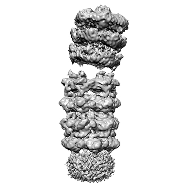 emd_0786.png emd_0786.png | 40.5 KB | ||
| Archive directory |  http://ftp.pdbj.org/pub/emdb/structures/EMD-0786ftp://ftp.pdbj.org/pub/emdb/structures/EMD-0786 http://ftp.pdbj.org/pub/emdb/structures/EMD-0786ftp://ftp.pdbj.org/pub/emdb/structures/EMD-0786 | HTTPS FTP |
-Related structure data
| Related structure data |  0784C  0785C  0787C  0788C  6kz3C  6kz4C C: citing same article ( |
|---|---|
| Similar structure data |
-Links
| EMDB pages | EMDB (EBI/PDBe) / EMDataResource |
|---|
-Map
| File | Download / File: emd_0786.map.gz / Format: CCP4 / Size: 178 MB / Type: IMAGE STORED AS FLOATING POINT NUMBER (4 BYTES) | ||||||||||||||||||||||||||||||||||||||||||||||||||||||||||||||||||||
|---|---|---|---|---|---|---|---|---|---|---|---|---|---|---|---|---|---|---|---|---|---|---|---|---|---|---|---|---|---|---|---|---|---|---|---|---|---|---|---|---|---|---|---|---|---|---|---|---|---|---|---|---|---|---|---|---|---|---|---|---|---|---|---|---|---|---|---|---|---|
| Annotation | The whole map of YebT | ||||||||||||||||||||||||||||||||||||||||||||||||||||||||||||||||||||
| Voxel size | X=Y=Z: 1.33 Å | ||||||||||||||||||||||||||||||||||||||||||||||||||||||||||||||||||||
| Density |
| ||||||||||||||||||||||||||||||||||||||||||||||||||||||||||||||||||||
| Symmetry | Space group: 1 | ||||||||||||||||||||||||||||||||||||||||||||||||||||||||||||||||||||
| Details | EMDB XML:
CCP4 map header:
| ||||||||||||||||||||||||||||||||||||||||||||||||||||||||||||||||||||
-Supplemental data
- Sample components
-Entire : YebT
| Entire | Name: YebT |
|---|---|
| Components |
|
-Supramolecule #1: YebT
| Supramolecule | Name: YebT / type: complex / ID: 1 / Parent: 0 / Macromolecule list: all / Details: Full length YebT |
|---|---|
| Source (natural) | Organism: Escherichia coli K-12 (bacteria) |
| Recombinant expression | Organism: Escherichia coli K-12 (bacteria) |
| Molecular weight | Theoretical: 570 KDa |
-Macromolecule #1: YebT
| Macromolecule | Name: YebT / type: protein_or_peptide / ID: 1 / Enantiomer: LEVO |
|---|---|
| Source (natural) | Organism: Escherichia coli K-12 (bacteria) |
| Sequence | String: MSQETPASTT EAQIKNKRRI SPFWLLPFIA LMIASWLIWD SYQDRGNTVT IDFMSADGIV PGRTPVRYQ GVEVGTVQDI SLSDDLRKIE VKVSIKSDMK DALREETQFW LVTPKASLAG V SGLDALVG GNYIGMMPGK GKEQDHFVAL DTQPKYRLDN GDLMIHLQAP ...String: MSQETPASTT EAQIKNKRRI SPFWLLPFIA LMIASWLIWD SYQDRGNTVT IDFMSADGIV PGRTPVRYQ GVEVGTVQDI SLSDDLRKIE VKVSIKSDMK DALREETQFW LVTPKASLAG V SGLDALVG GNYIGMMPGK GKEQDHFVAL DTQPKYRLDN GDLMIHLQAP DLGSLNSGSL VY FRKIPVG KVYDYAINPN KQGVVIDVLI ERRFTDLVKK GSRFWNVSGV DANVSISGAK VKL ESLAAL VNGAIAFDSP EESKPAEAED TFGLYEDLAH SQRGVIIKLE LPSGAGLTAD STPL MYQGL EVGQLTKLDL NPGGKVTGEM TVDPSVVTLL RENTRIELRN PKLSLSDANL SALLT GKTF ELVPGDGEPR KEFVVVPGEK ALLHEPDVLT LTLTAPESYG IDAGQPLILH GVQVGQ VID RKLTSKGVTF TVAIEPQHRE LVKGDSKFVV NSRVDVKVGL DGVEFLGASA SEWINGG IR ILPGDKGEMK ASYPLYANLE KALENSLSDL PTTTVSLSAE TLPDVQAGSV VLYRKFEV G EVITVRPRAN AFDIDLHIKP EYRNLLTSNS VFWAEGGAKV QLNGSGLTVQ ASPLSRALK GAISFDNLSG ASASQRKGDK RILYASETAA RAVGGQITLH AFDAGKLAVG MPIRYLGIDI GQIQTLDLI TARNEVQAKA VLYPEYVQTF ARGGTRFSVV TPQISAAGVE HLDTILQPYI N VEPGRGNP RRDFELQEAT ITDSRYLDGL SIIVEAPEAG SLGIGTPVLF RGLEVGTVTG MT LGTLSDR VMIAMRISKR YQHLVRNNSV FWLASGYSLD FGLTGGVVKT GTFNQFIRGG IAF ATPPGT PLAPKAQEGK HFLLQESEPK EWREWGTALP K |
-Experimental details
-Structure determination
| Method | cryo EM |
|---|---|
 Processing Processing | single particle reconstruction |
| Aggregation state | particle |
-Sample preparation
| Buffer | pH: 8 |
|---|---|
| Vitrification | Cryogen name: ETHANE |
- Electron microscopy
| Microscope | FEI TITAN KRIOS |
|---|---|
| Electron beam | Acceleration voltage: 300 kV / Electron source: FIELD EMISSION GUN |
| Electron optics | Illumination mode: FLOOD BEAM / Imaging mode: BRIGHT FIELDBright-field microscopy |
| Image recording | Film or detector model: GATAN K2 SUMMIT (4k x 4k) / Detector mode: SUPER-RESOLUTION / Average electron dose: 50.0 e/Å2 |
| Experimental equipment |  Model: Titan Krios / Image courtesy: FEI Company |
-Image processing
| Initial angle assignment | Type: MAXIMUM LIKELIHOOD |
|---|---|
| Final angle assignment | Type: MAXIMUM LIKELIHOOD |
| Final reconstruction | Applied symmetry - Point group: C1 (asymmetric) / Resolution.type: BY AUTHOR / Resolution: 4.6 Å / Resolution method: FSC 0.143 CUT-OFF / Number images used: 420134 |
| FSC plot (resolution estimation) |  |
