 Movie
Movie Controller

[English] 日本語
 Yorodumi
- PDB-2ca5: MxiH needle protein of Shigella Flexneri (monomeric form, residue... -
+ Open data

- Basic information
| Entry | Database: PDB / ID: 2ca5 | ||||||
|---|---|---|---|---|---|---|---|
| Title | MxiH needle protein of Shigella Flexneri (monomeric form, residues 1- 78) | ||||||
 Components Components | MXIH | ||||||
 Keywords Keywords |  TRANSPORT PROTEIN / MXIH / TYPE III SECRETION SYSTEM / NEEDLE COMPLEX / PROTEIN TRANSPORT / VIRULENCE TRANSPORT PROTEIN / MXIH / TYPE III SECRETION SYSTEM / NEEDLE COMPLEX / PROTEIN TRANSPORT / VIRULENCE | ||||||
| Function / homology |  Function and homology informationtype III protein secretion system complex / protein secretion by the type III secretion system / cell surface / extracellular region / identical protein binding Function and homology informationtype III protein secretion system complex / protein secretion by the type III secretion system / cell surface / extracellular region / identical protein bindingSimilarity search - Function | ||||||
| Biological species |  SHIGELLA FLEXNERI (bacteria) SHIGELLA FLEXNERI (bacteria) | ||||||
| Method | X-RAY DIFFRACTION / SYNCHROTRON / MIRAS / Resolution: 2.1 Å | ||||||
 Authors Authors | Deane, J.E. / Roversi, P. / Cordes, F.S. / Johnson, S. / Kenjale, R. / Picking, W.L. / Picking, W.D. / Blocker, A.J. / Lea, S.M. | ||||||
 Citation Citation | Journal: Proc Natl Acad Sci U S A / Year: 2006 Title: Molecular model of a type III secretion system needle: Implications for host-cell sensing. Authors: Janet E Deane / Pietro Roversi / Frank S Cordes / Steven Johnson / Roma Kenjale / Sarah Daniell / Frank Booy / William D Picking / Wendy L Picking / Ariel J Blocker / Susan M Lea /  Abstract: Type III secretion systems are essential virulence determinants for many Gram-negative bacterial pathogens. The type III secretion system consists of cytoplasmic, transmembrane, and extracellular ...Type III secretion systems are essential virulence determinants for many Gram-negative bacterial pathogens. The type III secretion system consists of cytoplasmic, transmembrane, and extracellular domains. The extracellular domain is a hollow needle protruding above the bacterial surface and is held within a basal body that traverses both bacterial membranes. Effector proteins are translocated, via this external needle, directly into host cells, where they subvert normal cell functions to aid infection. Physical contact with host cells initiates secretion and leads to formation of a pore, thought to be contiguous with the needle channel, in the host-cell membrane. Here, we report the crystal structure of the Shigella flexneri needle subunit MxiH and a complete model for the needle assembly built into our three-dimensional EM reconstruction. The model, combined with mutagenesis data, reveals that signaling of host-cell contact is relayed through the needle via intersubunit contacts and suggests a mode of binding for a tip complex. #1: Journal: Acta Crystallogr.,Sect.F / Year: 2006 Title: Expression, Purification, Crystallization and Preliminary Crystallographic Analysis of Mxih, a Subunit of the Shigella Flexneri Type III Secretion System Needle. Authors: Deane, J.E. / Cordes, F.S. / Roversi, P. / Johnson, S. / Kenjale, R. / Picking, W.D. / Picking, W.L. / Lea, S.M. / Blocker, A. | ||||||
| History |
|
- Structure visualization
| Structure viewer | Molecule: MolmilJmol/JSmol |
|---|
- Downloads & links
-Download
| PDBx/mmCIF format | 2ca5.cif.gz | 34.5 KB | Display | PDBx/mmCIF format |
|---|---|---|---|---|
| PDB format | pdb2ca5.ent.gz | 26.7 KB | Display | PDB format |
| PDBx/mmJSON format | 2ca5.json.gz | Tree view | PDBx/mmJSON format | |
| Others |  Other downloads Other downloads |
-Validation report
| Arichive directory | https://data.pdbj.org/pub/pdb/validation_reports/ca/2ca5ftp://data.pdbj.org/pub/pdb/validation_reports/ca/2ca5 | HTTPS FTP |
|---|
-Related structure data


-Links
 PDBj
PDBj
- Assembly
| Deposited unit | 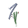
| ||||||||
|---|---|---|---|---|---|---|---|---|---|
| 1 | 
| ||||||||
| 2 | 
| ||||||||
| Unit cell |
| ||||||||
| Noncrystallographic symmetry (NCS) | NCS oper: (Code: given Matrix: (0.99021, 0.10552, 0.09133), Vector : |
-Components
| #1: Protein | Mass: 9545.514 Da / Num. of mol.: 2 / Fragment: TRUNCATED C-TERMINUS, RESIDUES 1-78 Source method: isolated from a genetically manipulated source Source: (gene. exp.) SHIGELLA FLEXNERI (bacteria) / Strain: PWR100 / Plasmid: PET22B / Production host: ESCHERICHIA COLI (E. coli) / Strain (production host): BL21(DE3) / References: UniProt: P0A223#2: Chemical | Isopropyl alcohol  Mass: 60.095 Da / Num. of mol.: 2 / Source method: obtained synthetically / Formula: C3H8O / Comment: alkaloid*YM Mass: 60.095 Da / Num. of mol.: 2 / Source method: obtained synthetically / Formula: C3H8O / Comment: alkaloid*YM#3: Chemical | ChemComp-GOL / | Glycerol  Mass: 92.094 Da / Num. of mol.: 1 / Source method: obtained synthetically / Formula: C3H8O3 Mass: 92.094 Da / Num. of mol.: 1 / Source method: obtained synthetically / Formula: C3H8O3#4: Chemical |   Mass: 22.990 Da / Num. of mol.: 2 / Source method: obtained synthetically / Formula: Na Mass: 22.990 Da / Num. of mol.: 2 / Source method: obtained synthetically / Formula: Na#5: Water | ChemComp-HOH / | Water Mass: 18.015 Da / Num. of mol.: 48 / Source method: isolated from a natural source / Formula: H2O Mass: 18.015 Da / Num. of mol.: 48 / Source method: isolated from a natural source / Formula: H2OSequence details | THIS CONSTRUCT IS A 5-RESIDUES C-TERMINAL DELETION MUTANT. THE LEHHHHH SEQUENCE COMES FROM THE ...THIS CONSTRUCT IS A 5-RESIDUES C-TERMINAL DELETION MUTANT. THE LEHHHHH SEQUENCE COMES FROM THE EXPRESSION | |
|---|
-Experimental details
-Experiment
| Experiment | Method: X-RAY DIFFRACTION / Number of used crystals: 1 |
|---|
- Sample preparation
| Crystal | Density Matthews: 1.9 Å3/Da / Density % sol: 0.34 % Description: STRUCTURE DETERMINED BY MIRAS WITH URANYL ACETATE AND SEMET DERIVATIVES |
|---|---|
| Crystal grow | pH: 5.6 Details: 20% PEG 4000, 0.1 M SODIUM CITRATE PH 5.6, 20% ISOPROPANOL, 10% GLYCEROL |
-Data collection
| Diffraction | Mean temperature: 100 K |
|---|---|
| Diffraction source | Source: SYNCHROTRON / Site: ESRF  / Beamline: ID14-4 / Wavelength: 0.9794 / Beamline: ID14-4 / Wavelength: 0.9794 |
| Detector | Type: ADSC CCD / Detector: CCD / Date: Jul 31, 2004 |
| Radiation | Protocol: SINGLE WAVELENGTH / Monochromatic (M) / Laue (L): M / Scattering type: x-ray |
| Radiation wavelength | Wavelength: 0.9794 Å / Relative weight: 1 |
| Reflection | Resolution: 2.1→28 Å / Num. obs: 8232 / % possible obs: 99.9 % / Observed criterion σ(I): 0 / Redundancy: 4.3 % / Biso Wilson estimate: 1.6 Å2 / Rmerge(I) obs: 0.07 / Net I/σ(I): 7.7 |
| Reflection shell | Resolution: 2.1→2.21 Å / Redundancy: 4.3 % / Rmerge(I) obs: 0.25 / Mean I/σ(I) obs: 2.9 / % possible all: 99.9 |
- Processing
| Software |
| |||||||||||||||||||||||||||||||||||||||||||||||||||||||||||||||||||||||||||||||||||||||||||||||
|---|---|---|---|---|---|---|---|---|---|---|---|---|---|---|---|---|---|---|---|---|---|---|---|---|---|---|---|---|---|---|---|---|---|---|---|---|---|---|---|---|---|---|---|---|---|---|---|---|---|---|---|---|---|---|---|---|---|---|---|---|---|---|---|---|---|---|---|---|---|---|---|---|---|---|---|---|---|---|---|---|---|---|---|---|---|---|---|---|---|---|---|---|---|---|---|---|
| Refinement | Method to determine structure: MIRAS / Resolution: 2.1→28 Å / Isotropic thermal model: TNT BCORREL / Cross valid method: THROUGHOUT / σ(F): 0 / Stereochemistry target values: TNT PROTGEO Details: REFINEMENT WAS DONE WITH MAXIMUM LIKELIHOOD IN BUSTER-TNT VERSION 1.3.0 THE REGIONS A1-A19, B1- B14 AND B76-B81 WERE DISORDERED IN THE CRYSTAL AND WERE NOT MODELLED
| |||||||||||||||||||||||||||||||||||||||||||||||||||||||||||||||||||||||||||||||||||||||||||||||
| Solvent computation | Solvent model: BABINET SCALING / Bsol: 25 Å2 / ksol: 0.418 e/Å3 | |||||||||||||||||||||||||||||||||||||||||||||||||||||||||||||||||||||||||||||||||||||||||||||||
| Refinement step | Cycle: LAST / Resolution: 2.1→28 Å
| |||||||||||||||||||||||||||||||||||||||||||||||||||||||||||||||||||||||||||||||||||||||||||||||
| Refine LS restraints |
|
